Iron »
PDB 3c25-3crb »
3c8f »
Iron in PDB 3c8f: 4FE-4S-Pyruvate Formate-Lyase Activating Enzyme with Partially Disordered Adomet
Enzymatic activity of 4FE-4S-Pyruvate Formate-Lyase Activating Enzyme with Partially Disordered Adomet
All present enzymatic activity of 4FE-4S-Pyruvate Formate-Lyase Activating Enzyme with Partially Disordered Adomet:
1.97.1.4;
1.97.1.4;
Protein crystallography data
The structure of 4FE-4S-Pyruvate Formate-Lyase Activating Enzyme with Partially Disordered Adomet, PDB code: 3c8f
was solved by
J.L.Vey,
C.L.Drennan,
with X-Ray Crystallography technique. A brief refinement statistics is given in the table below:
| Resolution Low / High (Å) | 30.90 / 2.25 |
| Space group | P 31 2 1 |
| Cell size a, b, c (Å), α, β, γ (°) | 58.051, 58.051, 117.466, 90.00, 90.00, 120.00 |
| R / Rfree (%) | 22.4 / 29.8 |
Iron Binding Sites:
The binding sites of Iron atom in the 4FE-4S-Pyruvate Formate-Lyase Activating Enzyme with Partially Disordered Adomet
(pdb code 3c8f). This binding sites where shown within
5.0 Angstroms radius around Iron atom.
In total 4 binding sites of Iron where determined in the 4FE-4S-Pyruvate Formate-Lyase Activating Enzyme with Partially Disordered Adomet, PDB code: 3c8f:
Jump to Iron binding site number: 1; 2; 3; 4;
In total 4 binding sites of Iron where determined in the 4FE-4S-Pyruvate Formate-Lyase Activating Enzyme with Partially Disordered Adomet, PDB code: 3c8f:
Jump to Iron binding site number: 1; 2; 3; 4;
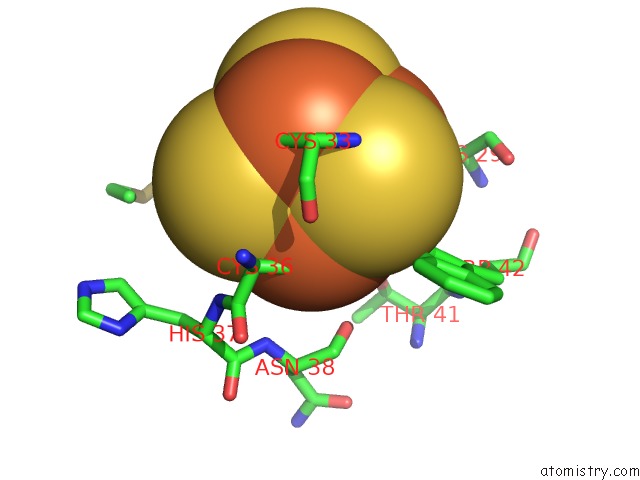
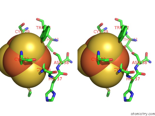
Iron binding site 1 out of 4 in 3c8f
Go back to
Iron binding site 1 out
of 4 in the 4FE-4S-Pyruvate Formate-Lyase Activating Enzyme with Partially Disordered Adomet
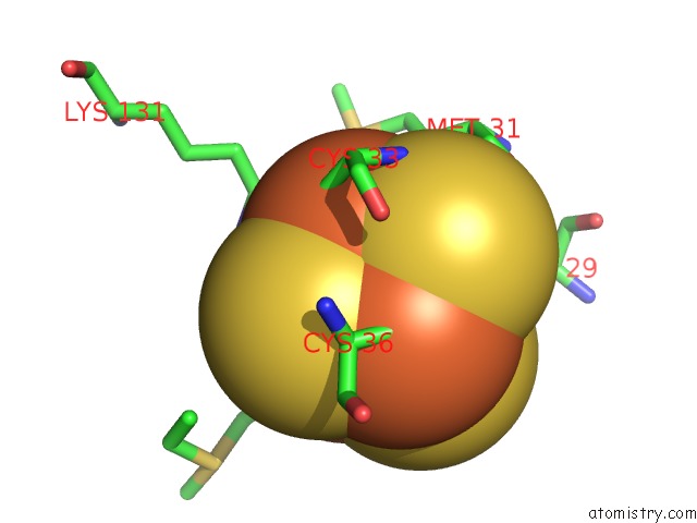
Mono view
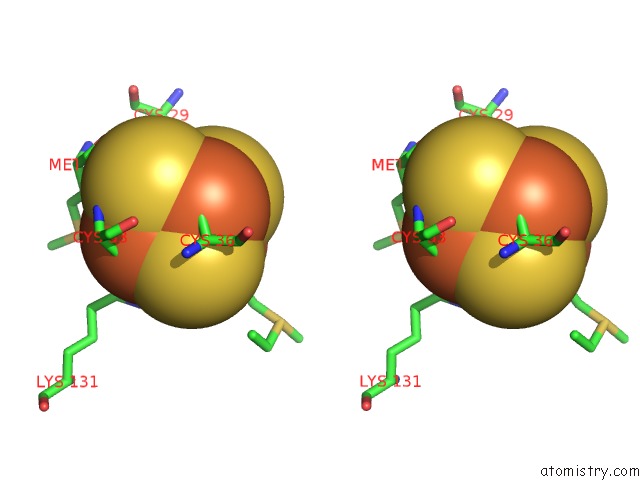
Stereo pair view
Mono view
Stereo pair view
A full contact list of Iron with other atoms in the Fe binding
site number 1 of 4FE-4S-Pyruvate Formate-Lyase Activating Enzyme with Partially Disordered Adomet within 5.0Å range:
|
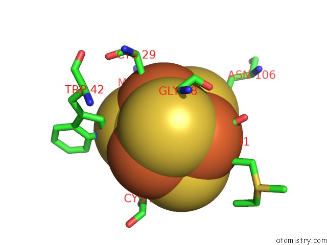
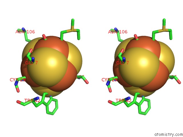
Iron binding site 2 out of 4 in 3c8f
Go back to
Iron binding site 2 out
of 4 in the 4FE-4S-Pyruvate Formate-Lyase Activating Enzyme with Partially Disordered Adomet
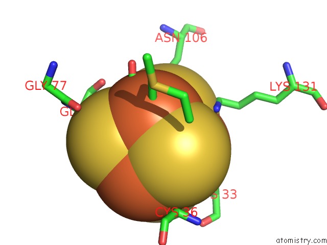
Mono view
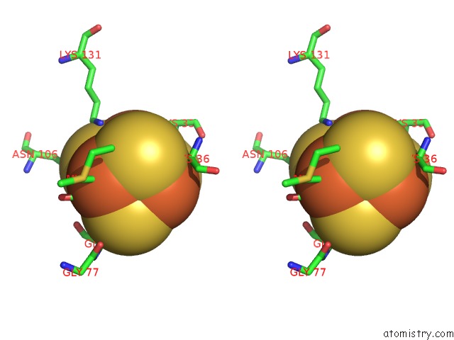
Stereo pair view
Mono view
Stereo pair view
A full contact list of Iron with other atoms in the Fe binding
site number 2 of 4FE-4S-Pyruvate Formate-Lyase Activating Enzyme with Partially Disordered Adomet within 5.0Å range:
|
Iron binding site 3 out of 4 in 3c8f
Go back to
Iron binding site 3 out
of 4 in the 4FE-4S-Pyruvate Formate-Lyase Activating Enzyme with Partially Disordered Adomet
Mono view
Stereo pair view
Mono view
Stereo pair view
A full contact list of Iron with other atoms in the Fe binding
site number 3 of 4FE-4S-Pyruvate Formate-Lyase Activating Enzyme with Partially Disordered Adomet within 5.0Å range:
|
Iron binding site 4 out of 4 in 3c8f
Go back to
Iron binding site 4 out
of 4 in the 4FE-4S-Pyruvate Formate-Lyase Activating Enzyme with Partially Disordered Adomet
Mono view
Stereo pair view
Mono view
Stereo pair view
A full contact list of Iron with other atoms in the Fe binding
site number 4 of 4FE-4S-Pyruvate Formate-Lyase Activating Enzyme with Partially Disordered Adomet within 5.0Å range:
|
Reference:
J.L.Vey,
J.Yang,
M.Li,
W.E.Broderick,
J.B.Broderick,
C.L.Drennan.
Structural Basis For Glycyl Radical Formation By Pyruvate Formate-Lyase Activating Enzyme. Proc.Natl.Acad.Sci.Usa V. 105 16137 2008.
ISSN: ISSN 0027-8424
PubMed: 18852451
DOI: 10.1073/PNAS.0806640105
Page generated: Sun Aug 4 08:15:10 2024
ISSN: ISSN 0027-8424
PubMed: 18852451
DOI: 10.1073/PNAS.0806640105
Last articles
F in 7MMHF in 7MMA
F in 7MM8
F in 7MM9
F in 7MHS
F in 7MII
F in 7MM7
F in 7MM6
F in 7MM5
F in 7MIG