Atomistry » Iron » PDB 3c25-3crb » 3chu
Atomistry »
  Iron »
    PDB 3c25-3crb »
      3chu »

Iron in PDB 3chu: Crystal Structure of Di-Iron Aurf

Protein crystallography data

The structure of Crystal Structure of Di-Iron Aurf, PDB code: 3chu was solved by H.Zhang, J.S.Brunzelle, S.K.Nair, with X-Ray Crystallography technique. A brief refinement statistics is given in the table below:

Resolution Low / High (Å) 25.00 / 2.20
Space group P 21 21 21
Cell size a, b, c (Å), α, β, γ (°) 57.979, 72.534, 138.792, 90.00, 90.00, 90.00
R / Rfree (%) 20.1 / 26.1

Iron Binding Sites:

The binding sites of Iron atom in the Crystal Structure of Di-Iron Aurf (pdb code 3chu). This binding sites where shown within 5.0 Angstroms radius around Iron atom.
In total 4 binding sites of Iron where determined in the Crystal Structure of Di-Iron Aurf, PDB code: 3chu:
Jump to Iron binding site number: 1; 2; 3; 4;

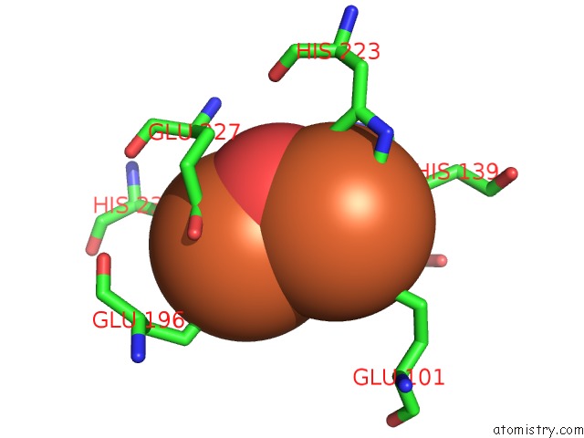
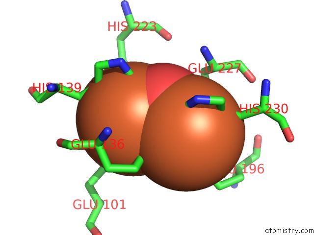
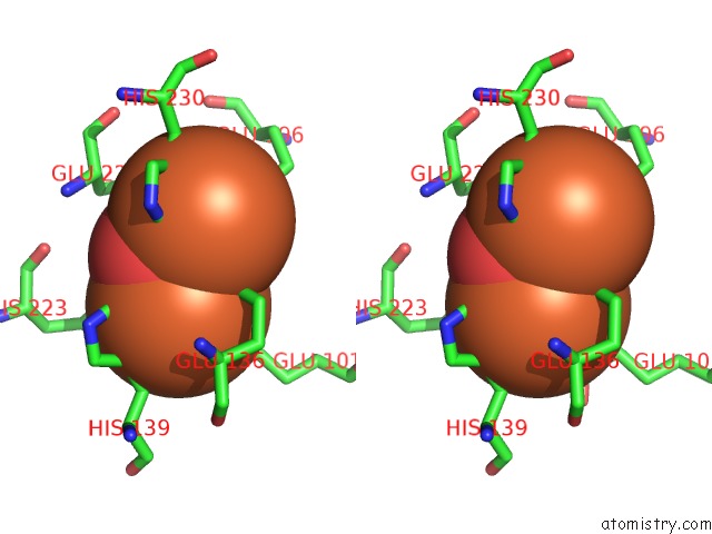
Iron binding site 1 out of 4 in 3chu

Go back to Iron Binding Sites List in 3chu
Iron binding site 1 out of 4 in the Crystal Structure of Di-Iron Aurf


Mono view


Stereo pair view

A full contact list of Iron with other atoms in the Fe binding site number 1 of Crystal Structure of Di-Iron Aurf within 5.0Å range:
probe atom residue distance (Å) B Occ
A:Fe501

b:25.2
occ:1.00
FE1 A:FEO501 0.0 25.2 1.0
OE1 A:GLU101 2.1 40.9 1.0
O A:FEO501 2.1 21.1 1.0
OE1 A:GLU136 2.2 24.4 1.0
ND1 A:HIS139 2.4 25.3 1.0
NE2 A:HIS223 2.4 27.1 1.0
CD A:GLU101 2.9 40.8 1.0
OE2 A:GLU101 3.2 43.1 1.0
CE1 A:HIS223 3.2 27.9 1.0
CD A:GLU136 3.2 24.4 1.0
CE1 A:HIS139 3.3 26.3 1.0
CD2 A:HIS223 3.3 28.6 1.0
CG A:HIS139 3.3 24.4 1.0
FE2 A:FEO501 3.5 24.1 1.0
CB A:HIS139 3.6 23.4 1.0
OE2 A:GLU136 3.6 27.1 1.0
ND1 A:HIS223 4.3 27.9 1.0
CG A:GLU101 4.3 36.0 1.0
CG A:HIS223 4.4 27.6 1.0
NE2 A:HIS139 4.4 26.6 1.0
CD2 A:HIS139 4.4 25.4 1.0
CA A:GLU136 4.4 24.4 1.0
OE2 A:GLU227 4.5 37.3 1.0
CG A:GLU136 4.5 24.7 1.0
OE2 A:GLU196 4.6 37.4 1.0
CB A:GLU136 4.8 23.5 1.0
ND1 A:HIS230 4.8 22.7 1.0
CE1 A:HIS230 4.9 20.4 1.0

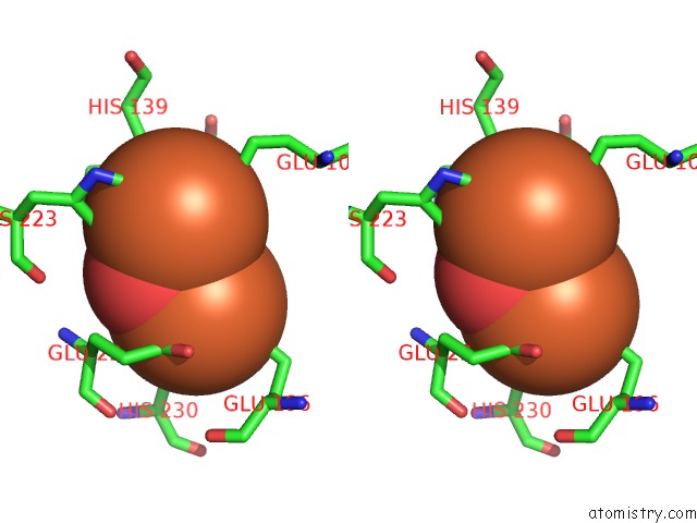
Iron binding site 2 out of 4 in 3chu

Go back to Iron Binding Sites List in 3chu
Iron binding site 2 out of 4 in the Crystal Structure of Di-Iron Aurf


Mono view


Stereo pair view

A full contact list of Iron with other atoms in the Fe binding site number 2 of Crystal Structure of Di-Iron Aurf within 5.0Å range:
probe atom residue distance (Å) B Occ
A:Fe501

b:24.1
occ:1.00
FE2 A:FEO501 0.0 24.1 1.0
O A:FEO501 2.0 21.1 1.0
OE1 A:GLU196 2.0 36.6 1.0
OE2 A:GLU136 2.1 27.1 1.0
OE2 A:GLU227 2.2 37.3 1.0
ND1 A:HIS230 2.3 22.7 1.0
OE2 A:GLU196 2.7 37.4 1.0
CD A:GLU196 2.7 35.7 1.0
CD A:GLU136 3.0 24.4 1.0
CE1 A:HIS230 3.2 20.4 1.0
CG A:HIS230 3.3 22.8 1.0
OE1 A:GLU136 3.3 24.4 1.0
CD A:GLU227 3.3 39.2 1.0
FE1 A:FEO501 3.5 25.2 1.0
CB A:HIS230 3.6 24.7 1.0
OE1 A:GLU227 3.9 44.5 1.0
CG A:GLU196 4.2 34.2 1.0
NE2 A:HIS230 4.3 19.2 1.0
CD2 A:HIS230 4.4 21.7 1.0
CG A:GLU136 4.4 24.7 1.0
CG A:GLU227 4.6 33.5 1.0
CA A:GLU227 4.6 28.4 1.0
CE2 A:PHE109 4.6 23.8 1.0
CB A:GLU227 4.7 28.6 1.0
ND1 A:HIS139 4.8 25.3 1.0
CE1 A:HIS139 4.8 26.3 1.0
OE2 A:GLU101 4.8 43.1 1.0
CB A:GLU196 4.9 33.0 1.0
CZ A:PHE109 4.9 24.8 1.0

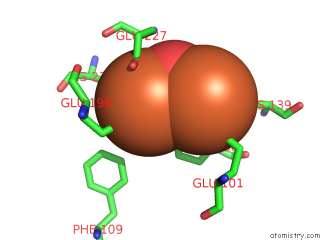
Iron binding site 3 out of 4 in 3chu

Go back to Iron Binding Sites List in 3chu
Iron binding site 3 out of 4 in the Crystal Structure of Di-Iron Aurf


Mono view


Stereo pair view

A full contact list of Iron with other atoms in the Fe binding site number 3 of Crystal Structure of Di-Iron Aurf within 5.0Å range:
probe atom residue distance (Å) B Occ
B:Fe501

b:20.0
occ:1.00
FE1 B:FEO501 0.0 20.0 1.0
OE2 B:GLU227 1.9 37.7 1.0
OE1 B:GLU196 2.0 28.2 1.0
OE2 B:GLU136 2.0 30.1 1.0
O B:FEO501 2.0 14.8 1.0
ND1 B:HIS230 2.1 22.1 1.0
CD B:GLU196 2.7 32.4 1.0
OE2 B:GLU196 2.7 31.1 1.0
CD B:GLU136 3.0 27.6 1.0
CD B:GLU227 3.0 37.8 1.0
CE1 B:HIS230 3.1 23.4 1.0
CG B:HIS230 3.2 27.2 1.0
OE1 B:GLU136 3.3 23.4 1.0
OE1 B:GLU227 3.5 43.0 1.0
FE2 B:FEO501 3.5 19.6 1.0
CB B:HIS230 3.6 26.7 1.0
CG B:GLU196 4.1 30.3 1.0
NE2 B:HIS230 4.2 25.8 1.0
CD2 B:HIS230 4.3 24.5 1.0
CG B:GLU227 4.3 32.8 1.0
CG B:GLU136 4.4 26.9 1.0
CA B:GLU227 4.5 28.1 1.0
CE1 B:HIS139 4.7 26.8 1.0
ND1 B:HIS139 4.7 26.4 1.0
CE2 B:PHE109 4.7 31.5 1.0
CB B:GLU227 4.8 29.1 1.0
CB B:GLU196 4.8 29.8 1.0
CZ B:PHE109 4.9 32.0 1.0

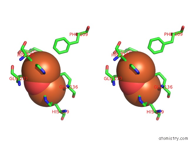
Iron binding site 4 out of 4 in 3chu

Go back to Iron Binding Sites List in 3chu
Iron binding site 4 out of 4 in the Crystal Structure of Di-Iron Aurf


Mono view


Stereo pair view

A full contact list of Iron with other atoms in the Fe binding site number 4 of Crystal Structure of Di-Iron Aurf within 5.0Å range:
probe atom residue distance (Å) B Occ
B:Fe501

b:19.6
occ:1.00
FE2 B:FEO501 0.0 19.6 1.0
O B:FEO501 1.9 14.8 1.0
OE1 B:GLU136 2.0 23.4 1.0
OE1 B:GLU101 2.1 40.1 1.0
ND1 B:HIS139 2.3 26.4 1.0
NE2 B:HIS223 2.3 30.9 1.0
CD B:GLU136 3.1 27.6 1.0
CD B:GLU101 3.1 39.0 1.0
CD2 B:HIS223 3.1 29.3 1.0
CE1 B:HIS139 3.2 26.8 1.0
CE1 B:HIS223 3.3 31.3 1.0
CG B:HIS139 3.3 25.9 1.0
OE2 B:GLU136 3.5 30.1 1.0
FE1 B:FEO501 3.5 20.0 1.0
CB B:HIS139 3.6 25.7 1.0
CG B:GLU101 3.7 36.3 1.0
OE1 B:GLU227 4.0 43.0 1.0
OE2 B:GLU227 4.1 37.7 1.0
OE2 B:GLU101 4.1 41.5 1.0
CG B:HIS223 4.3 29.2 1.0
ND1 B:HIS223 4.3 31.9 1.0
NE2 B:HIS139 4.3 27.6 1.0
CD B:GLU227 4.3 37.8 1.0
CD2 B:HIS139 4.4 25.8 1.0
CG B:GLU136 4.4 26.9 1.0
CA B:GLU136 4.5 27.1 1.0
OE2 B:GLU196 4.6 31.1 1.0
CB B:GLU136 4.7 26.2 1.0
ND1 B:HIS230 4.8 22.1 1.0
CE1 B:HIS230 4.8 23.4 1.0

Reference:

Y.S.Choi, H.Zhang, J.S.Brunzelle, S.K.Nair, H.Zhao. In Vitro Reconstitution and Crystal Structure of P-Aminobenzoate N-Oxygenase (Aurf) Involved in Aureothin Biosynthesis. Proc.Natl.Acad.Sci.Usa V. 105 6858 2008.
ISSN: ISSN 0027-8424
PubMed: 18458342
DOI: 10.1073/PNAS.0712073105
Page generated: Sun Dec 13 15:02:38 2020

Last articles

Zn in 8WB0
Zn in 8WAX
Zn in 8WAU
Zn in 8WAZ
Zn in 8WAY
Zn in 8WAV
Zn in 8WAW
Zn in 8WAT
Zn in 8W7M
Zn in 8WD3
© Copyright 2008-2020 by atomistry.com
Home   |    Site Map   |    Copyright   |    Contact us   |    Privacy