Iron »
PDB 3crv-3dby »
3crv »
Iron in PDB 3crv: XPD_HELICASE
Protein crystallography data
The structure of XPD_HELICASE, PDB code: 3crv
was solved by
L.Fan,
A.S.Arvai,
J.A.Tainer,
with X-Ray Crystallography technique. A brief refinement statistics is given in the table below:
| Resolution Low / High (Å) | 42.58 / 2.00 |
| Space group | P 21 21 21 |
| Cell size a, b, c (Å), α, β, γ (°) | 53.542, 70.222, 144.293, 90.00, 90.00, 90.00 |
| R / Rfree (%) | 22.2 / 26 |
Iron Binding Sites:
The binding sites of Iron atom in the XPD_HELICASE
(pdb code 3crv). This binding sites where shown within
5.0 Angstroms radius around Iron atom.
In total 4 binding sites of Iron where determined in the XPD_HELICASE, PDB code: 3crv:
Jump to Iron binding site number: 1; 2; 3; 4;
In total 4 binding sites of Iron where determined in the XPD_HELICASE, PDB code: 3crv:
Jump to Iron binding site number: 1; 2; 3; 4;
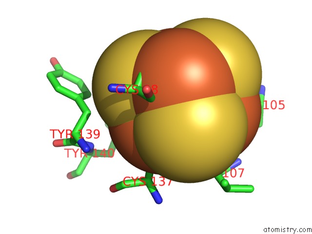
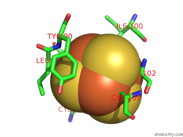
Iron binding site 1 out of 4 in 3crv
Go back to
Iron binding site 1 out
of 4 in the XPD_HELICASE
Mono view
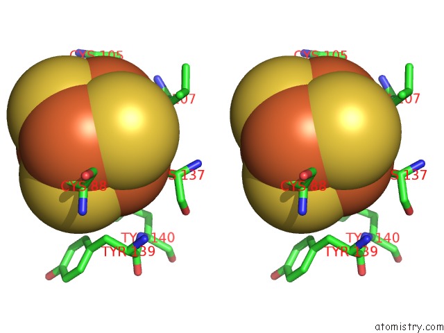
Stereo pair view
Mono view
Stereo pair view
A full contact list of Iron with other atoms in the Fe binding
site number 1 of XPD_HELICASE within 5.0Å range:
|
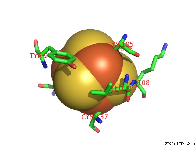
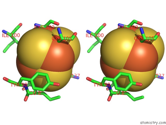
Iron binding site 2 out of 4 in 3crv
Go back to
Iron binding site 2 out
of 4 in the XPD_HELICASE
Mono view
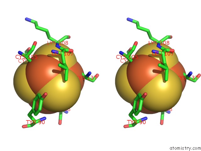
Stereo pair view
Mono view
Stereo pair view
A full contact list of Iron with other atoms in the Fe binding
site number 2 of XPD_HELICASE within 5.0Å range:
|
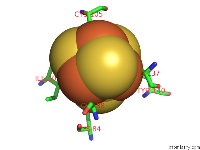
Iron binding site 3 out of 4 in 3crv
Go back to
Iron binding site 3 out
of 4 in the XPD_HELICASE
Mono view
Stereo pair view
Mono view
Stereo pair view
A full contact list of Iron with other atoms in the Fe binding
site number 3 of XPD_HELICASE within 5.0Å range:
|
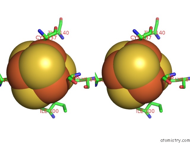
Iron binding site 4 out of 4 in 3crv
Go back to
Iron binding site 4 out
of 4 in the XPD_HELICASE
Mono view
Stereo pair view
Mono view
Stereo pair view
A full contact list of Iron with other atoms in the Fe binding
site number 4 of XPD_HELICASE within 5.0Å range:
|
Reference:
L.Fan,
J.O.Fuss,
Q.J.Cheng,
A.S.Arvai,
M.Hammel,
V.A.Roberts,
P.K.Cooper,
J.A.Tainer.
Xpd Helicase Structures and Activities: Insights Into the Cancer and Aging Phenotypes From Xpd Mutations. Cell(Cambridge,Mass.) V. 133 789 2008.
ISSN: ISSN 0092-8674
PubMed: 18510924
DOI: 10.1016/J.CELL.2008.04.030
Page generated: Sun Aug 4 08:33:46 2024
ISSN: ISSN 0092-8674
PubMed: 18510924
DOI: 10.1016/J.CELL.2008.04.030
Last articles
Zn in 9MJ5Zn in 9HNW
Zn in 9G0L
Zn in 9FNE
Zn in 9DZN
Zn in 9E0I
Zn in 9D32
Zn in 9DAK
Zn in 8ZXC
Zn in 8ZUF