Iron »
PDB 3crv-3dby »
3cyt »
Iron in PDB 3cyt: Redox Conformation Changes in Refined Tuna Cytochrome C
Protein crystallography data
The structure of Redox Conformation Changes in Refined Tuna Cytochrome C, PDB code: 3cyt
was solved by
T.Takano,
with X-Ray Crystallography technique. A brief refinement statistics is given in the table below:
| Resolution Low / High (Å) | N/A / 1.80 |
| Space group | P 43 |
| Cell size a, b, c (Å), α, β, γ (°) | 74.420, 74.420, 36.300, 90.00, 90.00, 90.00 |
| R / Rfree (%) | n/a / n/a |
Iron Binding Sites:
The binding sites of Iron atom in the Redox Conformation Changes in Refined Tuna Cytochrome C
(pdb code 3cyt). This binding sites where shown within
5.0 Angstroms radius around Iron atom.
In total 2 binding sites of Iron where determined in the Redox Conformation Changes in Refined Tuna Cytochrome C, PDB code: 3cyt:
Jump to Iron binding site number: 1; 2;
In total 2 binding sites of Iron where determined in the Redox Conformation Changes in Refined Tuna Cytochrome C, PDB code: 3cyt:
Jump to Iron binding site number: 1; 2;
Iron binding site 1 out of 2 in 3cyt
Go back to
Iron binding site 1 out
of 2 in the Redox Conformation Changes in Refined Tuna Cytochrome C
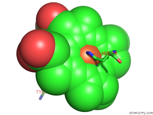
Mono view
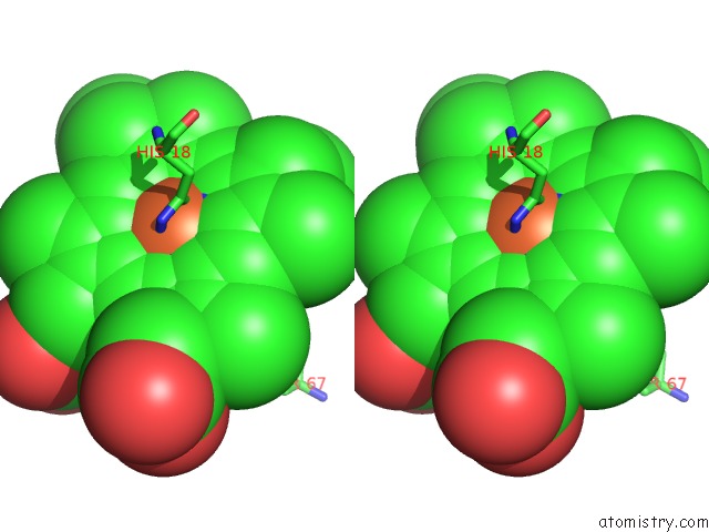
Stereo pair view
Mono view
Stereo pair view
A full contact list of Iron with other atoms in the Fe binding
site number 1 of Redox Conformation Changes in Refined Tuna Cytochrome C within 5.0Å range:
|
Iron binding site 2 out of 2 in 3cyt
Go back to
Iron binding site 2 out
of 2 in the Redox Conformation Changes in Refined Tuna Cytochrome C
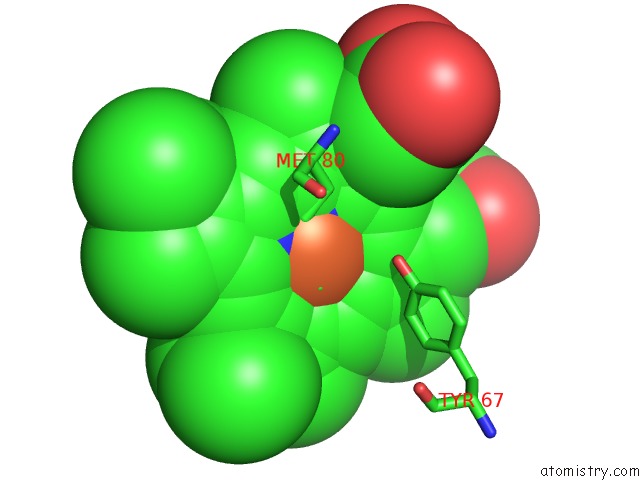
Mono view
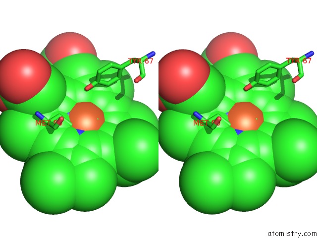
Stereo pair view
Mono view
Stereo pair view
A full contact list of Iron with other atoms in the Fe binding
site number 2 of Redox Conformation Changes in Refined Tuna Cytochrome C within 5.0Å range:
|
Reference:
T.Takano,
R.E.Dickerson.
Redox Conformation Changes in Refined Tuna Cytochrome C. Proc.Natl.Acad.Sci.Usa V. 77 6371 1980.
ISSN: ISSN 0027-8424
PubMed: 6256733
DOI: 10.1073/PNAS.77.11.6371
Page generated: Sun Aug 4 08:38:44 2024
ISSN: ISSN 0027-8424
PubMed: 6256733
DOI: 10.1073/PNAS.77.11.6371
Last articles
Zn in 9MJ5Zn in 9HNW
Zn in 9G0L
Zn in 9FNE
Zn in 9DZN
Zn in 9E0I
Zn in 9D32
Zn in 9DAK
Zn in 8ZXC
Zn in 8ZUF