Iron »
PDB 3crv-3dby »
3d1k »
Iron in PDB 3d1k: R/T Intermediate Quaternary Structure of An Antarctic Fish Hemoglobin in An Alpha(Co)-Beta(Pentacoordinate) State
Protein crystallography data
The structure of R/T Intermediate Quaternary Structure of An Antarctic Fish Hemoglobin in An Alpha(Co)-Beta(Pentacoordinate) State, PDB code: 3d1k
was solved by
L.Vitagliano,
A.Vergara,
G.Bonomi,
A.Merlino,
L.Mazzarella,
with X-Ray Crystallography technique. A brief refinement statistics is given in the table below:
| Resolution Low / High (Å) | 30.00 / 1.25 |
| Space group | C 1 2 1 |
| Cell size a, b, c (Å), α, β, γ (°) | 88.458, 87.664, 55.502, 90.00, 99.16, 90.00 |
| R / Rfree (%) | 16.7 / 20.2 |
Iron Binding Sites:
The binding sites of Iron atom in the R/T Intermediate Quaternary Structure of An Antarctic Fish Hemoglobin in An Alpha(Co)-Beta(Pentacoordinate) State
(pdb code 3d1k). This binding sites where shown within
5.0 Angstroms radius around Iron atom.
In total 2 binding sites of Iron where determined in the R/T Intermediate Quaternary Structure of An Antarctic Fish Hemoglobin in An Alpha(Co)-Beta(Pentacoordinate) State, PDB code: 3d1k:
Jump to Iron binding site number: 1; 2;
In total 2 binding sites of Iron where determined in the R/T Intermediate Quaternary Structure of An Antarctic Fish Hemoglobin in An Alpha(Co)-Beta(Pentacoordinate) State, PDB code: 3d1k:
Jump to Iron binding site number: 1; 2;
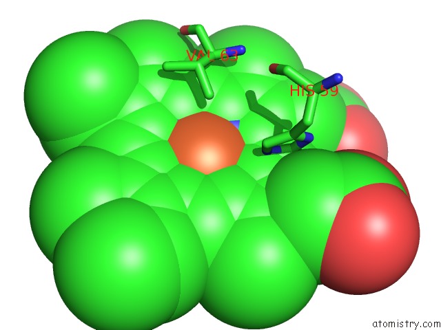
Iron binding site 1 out of 2 in 3d1k
Go back to
Iron binding site 1 out
of 2 in the R/T Intermediate Quaternary Structure of An Antarctic Fish Hemoglobin in An Alpha(Co)-Beta(Pentacoordinate) State
Mono view

Stereo pair view
Mono view
Stereo pair view
A full contact list of Iron with other atoms in the Fe binding
site number 1 of R/T Intermediate Quaternary Structure of An Antarctic Fish Hemoglobin in An Alpha(Co)-Beta(Pentacoordinate) State within 5.0Å range:
|
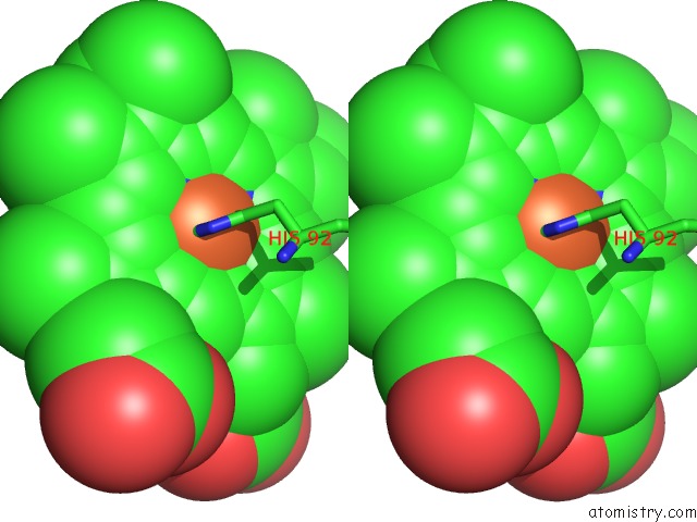
Iron binding site 2 out of 2 in 3d1k
Go back to
Iron binding site 2 out
of 2 in the R/T Intermediate Quaternary Structure of An Antarctic Fish Hemoglobin in An Alpha(Co)-Beta(Pentacoordinate) State

Mono view
Stereo pair view
Mono view
Stereo pair view
A full contact list of Iron with other atoms in the Fe binding
site number 2 of R/T Intermediate Quaternary Structure of An Antarctic Fish Hemoglobin in An Alpha(Co)-Beta(Pentacoordinate) State within 5.0Å range:
|
Reference:
L.Vitagliano,
A.Vergara,
G.Bonomi,
A.Merlino,
C.Verde,
G.Di Prisco,
B.D.Howes,
G.Smulevich,
L.Mazzarella.
Spectroscopic and Crystallographic Characterization of A Tetrameric Hemoglobin Oxidation Reveals Structural Features of the Functional Intermediate Relaxed/Tense State. J.Am.Chem.Soc. V. 130 10527 2008.
ISSN: ISSN 0002-7863
PubMed: 18642904
DOI: 10.1021/JA803363P
Page generated: Sun Aug 4 08:44:38 2024
ISSN: ISSN 0002-7863
PubMed: 18642904
DOI: 10.1021/JA803363P
Last articles
Zn in 9JYWZn in 9IR4
Zn in 9IR3
Zn in 9GMX
Zn in 9GMW
Zn in 9JEJ
Zn in 9ERF
Zn in 9ERE
Zn in 9EGV
Zn in 9EGW