Iron »
PDB 3crv-3dby »
3d89 »
Iron in PDB 3d89: Crystal Structure of A Soluble Rieske Ferredoxin From Mus Musculus
Protein crystallography data
The structure of Crystal Structure of A Soluble Rieske Ferredoxin From Mus Musculus, PDB code: 3d89
was solved by
E.J.Levin,
J.G.Mccoy,
N.L.Elsen,
K.D.Seder,
C.A.Bingman,
G.E.Wesenberg,
B.G.Fox,
G.N.Phillips Jr.,
Center For Eukaryotic Structural Genomics(Cesg),
with X-Ray Crystallography technique. A brief refinement statistics is given in the table below:
| Resolution Low / High (Å) | 37.75 / 2.07 |
| Space group | P 43 21 2 |
| Cell size a, b, c (Å), α, β, γ (°) | 52.413, 52.413, 108.808, 90.00, 90.00, 90.00 |
| R / Rfree (%) | 19.9 / 22.8 |
Iron Binding Sites:
The binding sites of Iron atom in the Crystal Structure of A Soluble Rieske Ferredoxin From Mus Musculus
(pdb code 3d89). This binding sites where shown within
5.0 Angstroms radius around Iron atom.
In total 2 binding sites of Iron where determined in the Crystal Structure of A Soluble Rieske Ferredoxin From Mus Musculus, PDB code: 3d89:
Jump to Iron binding site number: 1; 2;
In total 2 binding sites of Iron where determined in the Crystal Structure of A Soluble Rieske Ferredoxin From Mus Musculus, PDB code: 3d89:
Jump to Iron binding site number: 1; 2;
Iron binding site 1 out of 2 in 3d89
Go back to
Iron binding site 1 out
of 2 in the Crystal Structure of A Soluble Rieske Ferredoxin From Mus Musculus
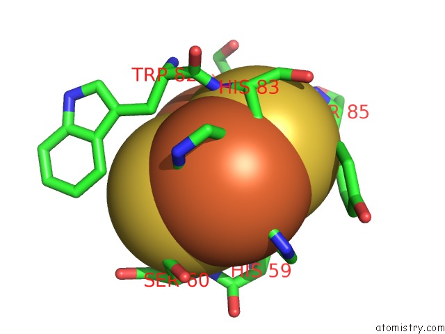
Mono view
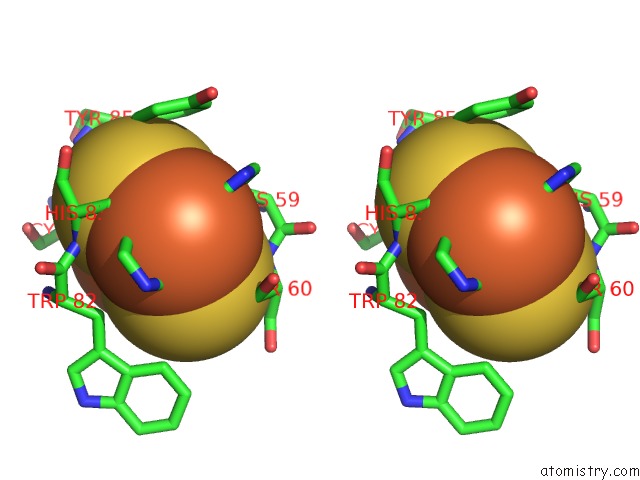
Stereo pair view
Mono view
Stereo pair view
A full contact list of Iron with other atoms in the Fe binding
site number 1 of Crystal Structure of A Soluble Rieske Ferredoxin From Mus Musculus within 5.0Å range:
|
Iron binding site 2 out of 2 in 3d89
Go back to
Iron binding site 2 out
of 2 in the Crystal Structure of A Soluble Rieske Ferredoxin From Mus Musculus
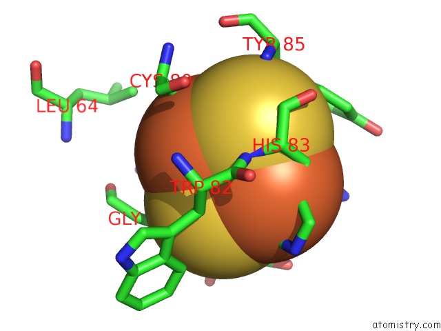
Mono view
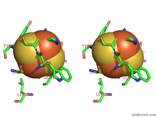
Stereo pair view
Mono view
Stereo pair view
A full contact list of Iron with other atoms in the Fe binding
site number 2 of Crystal Structure of A Soluble Rieske Ferredoxin From Mus Musculus within 5.0Å range:
|
Reference:
E.J.Levin,
N.L.Elsen,
K.D.Seder,
J.G.Mccoy,
B.G.Fox,
G.N.Phillips.
X-Ray Structure of A Soluble Rieske-Type Ferredoxin From Mus Musculus. Acta Crystallogr.,Sect.D V. 64 933 2008.
ISSN: ISSN 0907-4449
PubMed: 18703841
DOI: 10.1107/S0907444908021653
Page generated: Sun Aug 4 08:47:13 2024
ISSN: ISSN 0907-4449
PubMed: 18703841
DOI: 10.1107/S0907444908021653
Last articles
Zn in 9J0NZn in 9J0O
Zn in 9J0P
Zn in 9FJX
Zn in 9EKB
Zn in 9C0F
Zn in 9CAH
Zn in 9CH0
Zn in 9CH3
Zn in 9CH1