Iron »
PDB 3crv-3dby »
3daf »
Iron in PDB 3daf: The Crystal Structure of [Fe]-Hydrogenase Holoenzyme (Hmd) From Methanocaldococcus Jannaschii Cocrystallized with Cyanide
Enzymatic activity of The Crystal Structure of [Fe]-Hydrogenase Holoenzyme (Hmd) From Methanocaldococcus Jannaschii Cocrystallized with Cyanide
All present enzymatic activity of The Crystal Structure of [Fe]-Hydrogenase Holoenzyme (Hmd) From Methanocaldococcus Jannaschii Cocrystallized with Cyanide:
1.12.98.2;
1.12.98.2;
Protein crystallography data
The structure of The Crystal Structure of [Fe]-Hydrogenase Holoenzyme (Hmd) From Methanocaldococcus Jannaschii Cocrystallized with Cyanide, PDB code: 3daf
was solved by
O.Pilak,
E.Warkentin,
S.Shima,
R.K.Thauer,
U.Ermler,
with X-Ray Crystallography technique. A brief refinement statistics is given in the table below:
| Resolution Low / High (Å) | 10.00 / 1.75 |
| Space group | I 41 2 2 |
| Cell size a, b, c (Å), α, β, γ (°) | 97.140, 97.140, 166.030, 90.00, 90.00, 90.00 |
| R / Rfree (%) | 18.7 / 22.2 |
Iron Binding Sites:
The binding sites of Iron atom in the The Crystal Structure of [Fe]-Hydrogenase Holoenzyme (Hmd) From Methanocaldococcus Jannaschii Cocrystallized with Cyanide
(pdb code 3daf). This binding sites where shown within
5.0 Angstroms radius around Iron atom.
In total only one binding site of Iron was determined in the The Crystal Structure of [Fe]-Hydrogenase Holoenzyme (Hmd) From Methanocaldococcus Jannaschii Cocrystallized with Cyanide, PDB code: 3daf:
In total only one binding site of Iron was determined in the The Crystal Structure of [Fe]-Hydrogenase Holoenzyme (Hmd) From Methanocaldococcus Jannaschii Cocrystallized with Cyanide, PDB code: 3daf:
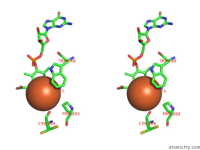
Iron binding site 1 out of 1 in 3daf
Go back to
Iron binding site 1 out
of 1 in the The Crystal Structure of [Fe]-Hydrogenase Holoenzyme (Hmd) From Methanocaldococcus Jannaschii Cocrystallized with Cyanide
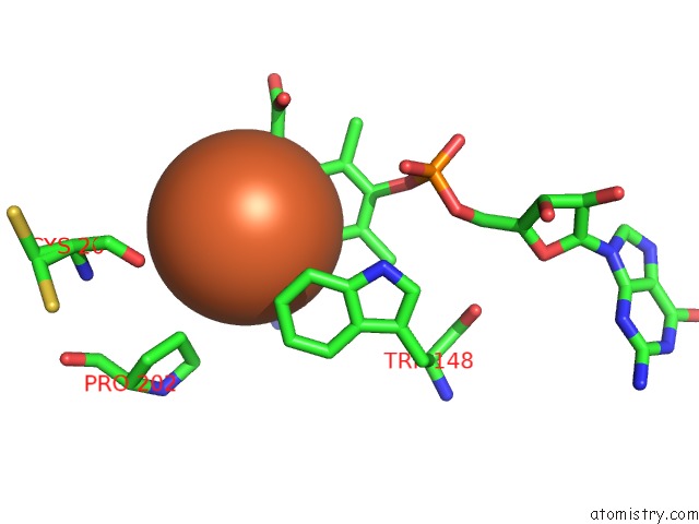
Mono view
Stereo pair view
Mono view
Stereo pair view
A full contact list of Iron with other atoms in the Fe binding
site number 1 of The Crystal Structure of [Fe]-Hydrogenase Holoenzyme (Hmd) From Methanocaldococcus Jannaschii Cocrystallized with Cyanide within 5.0Å range:
|
Reference:
S.Shima,
O.Pilak,
S.Vogt,
M.Schick,
M.S.Stagni,
W.Meyer-Klaucke,
E.Warkentin,
R.K.Thauer,
U.Ermler.
The Crystal Structure of [Fe]-Hydrogenase Reveals the Geometry of the Active Site. Science V. 321 572 2008.
ISSN: ISSN 0036-8075
PubMed: 18653896
DOI: 10.1126/SCIENCE.1158978
Page generated: Sun Aug 4 08:47:28 2024
ISSN: ISSN 0036-8075
PubMed: 18653896
DOI: 10.1126/SCIENCE.1158978
Last articles
Cl in 2X5OCl in 2X5K
Cl in 2X5G
Cl in 2X2J
Cl in 2X21
Cl in 2X3O
Cl in 2X3C
Cl in 2X39
Cl in 2X2H
Cl in 2X1D