Iron in PDB 3dhg: Crystal Structure of Toluene 4-Monoxygenase Hydroxylase
Protein crystallography data
The structure of Crystal Structure of Toluene 4-Monoxygenase Hydroxylase, PDB code: 3dhg
was solved by
L.J.Bailey,
J.G.Mccoy,
G.N.Phillips Jr.,
B.G.Fox,
with X-Ray Crystallography technique. A brief refinement statistics is given in the table below:
Resolution Low / High (Å)
|
49.45 /
1.85
|
Space group
|
P 1 21 1
|
Cell size a, b, c (Å), α, β, γ (°)
|
56.822,
181.481,
89.917,
90.00,
107.62,
90.00
|
R / Rfree (%)
|
16.4 /
21.4
|
Other elements in 3dhg:
The structure of Crystal Structure of Toluene 4-Monoxygenase Hydroxylase also contains other interesting chemical elements:
Iron Binding Sites:
The binding sites of Iron atom in the Crystal Structure of Toluene 4-Monoxygenase Hydroxylase
(pdb code 3dhg). This binding sites where shown within
5.0 Angstroms radius around Iron atom.
In total 4 binding sites of Iron where determined in the
Crystal Structure of Toluene 4-Monoxygenase Hydroxylase, PDB code: 3dhg:
Jump to Iron binding site number:
1;
2;
3;
4;
Iron binding site 1 out
of 4 in 3dhg
Go back to
Iron Binding Sites List in 3dhg
Iron binding site 1 out
of 4 in the Crystal Structure of Toluene 4-Monoxygenase Hydroxylase
Mono view
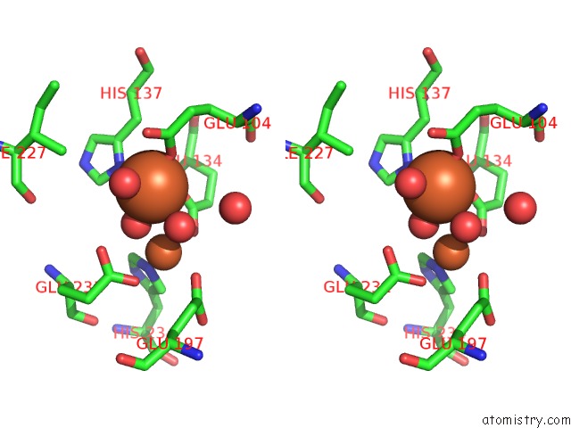
Stereo pair view
|
A full contact list of Iron with other atoms in the Fe binding
site number 1 of Crystal Structure of Toluene 4-Monoxygenase Hydroxylase within 5.0Å range:
probe
|
atom
|
residue
|
distance (Å)
|
B
|
Occ
|
A:Fe501
b:20.6
occ:1.00
|
O
|
A:HOH504
|
2.0
|
23.0
|
1.0
|
OE1
|
A:GLU104
|
2.0
|
20.8
|
1.0
|
OE1
|
A:GLU134
|
2.0
|
17.6
|
1.0
|
O
|
A:HOH506
|
2.1
|
19.0
|
1.0
|
ND1
|
A:HIS137
|
2.3
|
17.4
|
1.0
|
O
|
A:HOH505
|
2.7
|
31.8
|
1.0
|
CD
|
A:GLU134
|
3.0
|
19.4
|
1.0
|
CD
|
A:GLU104
|
3.1
|
22.4
|
1.0
|
CE1
|
A:HIS137
|
3.2
|
20.1
|
1.0
|
FE
|
A:FE502
|
3.3
|
23.1
|
1.0
|
CG
|
A:HIS137
|
3.3
|
19.0
|
1.0
|
OE2
|
A:GLU134
|
3.4
|
20.5
|
1.0
|
OE2
|
A:GLU104
|
3.5
|
22.2
|
1.0
|
CB
|
A:HIS137
|
3.6
|
17.2
|
1.0
|
OE1
|
A:GLU231
|
4.0
|
37.7
|
1.0
|
NE2
|
A:HIS137
|
4.4
|
19.3
|
1.0
|
CG
|
A:GLU104
|
4.4
|
20.5
|
1.0
|
CG
|
A:GLU134
|
4.4
|
16.0
|
1.0
|
CD2
|
A:HIS137
|
4.4
|
14.9
|
1.0
|
OE2
|
A:GLU231
|
4.7
|
33.5
|
1.0
|
NE2
|
A:HIS234
|
4.7
|
22.8
|
1.0
|
CB
|
A:GLU104
|
4.7
|
18.4
|
1.0
|
CA
|
A:GLU134
|
4.7
|
15.8
|
1.0
|
CG2
|
A:ILE227
|
4.7
|
18.1
|
1.0
|
CD
|
A:GLU231
|
4.8
|
31.6
|
1.0
|
CB
|
A:GLU134
|
4.8
|
15.1
|
1.0
|
CE1
|
A:HIS234
|
4.9
|
17.7
|
1.0
|
O
|
A:HOH1410
|
4.9
|
45.7
|
1.0
|
OE2
|
A:GLU197
|
4.9
|
25.2
|
1.0
|
|
Iron binding site 2 out
of 4 in 3dhg
Go back to
Iron Binding Sites List in 3dhg
Iron binding site 2 out
of 4 in the Crystal Structure of Toluene 4-Monoxygenase Hydroxylase
Mono view
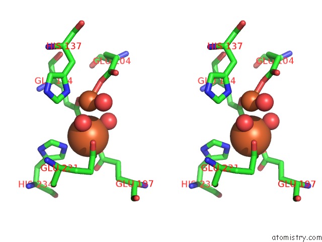
Stereo pair view
|
A full contact list of Iron with other atoms in the Fe binding
site number 2 of Crystal Structure of Toluene 4-Monoxygenase Hydroxylase within 5.0Å range:
probe
|
atom
|
residue
|
distance (Å)
|
B
|
Occ
|
A:Fe502
b:23.1
occ:1.00
|
O
|
A:HOH504
|
1.9
|
23.0
|
1.0
|
OE2
|
A:GLU197
|
2.0
|
25.2
|
1.0
|
OE2
|
A:GLU134
|
2.1
|
20.5
|
1.0
|
O
|
A:HOH505
|
2.2
|
31.8
|
1.0
|
OE2
|
A:GLU231
|
2.2
|
33.5
|
1.0
|
NE2
|
A:HIS234
|
2.2
|
22.8
|
1.0
|
CD
|
A:GLU197
|
3.0
|
21.9
|
1.0
|
CD
|
A:GLU134
|
3.1
|
19.4
|
1.0
|
CD
|
A:GLU231
|
3.1
|
31.6
|
1.0
|
CD2
|
A:HIS234
|
3.2
|
19.9
|
1.0
|
CE1
|
A:HIS234
|
3.2
|
17.7
|
1.0
|
FE
|
A:FE501
|
3.3
|
20.6
|
1.0
|
OE1
|
A:GLU231
|
3.3
|
37.7
|
1.0
|
OE1
|
A:GLU134
|
3.3
|
17.6
|
1.0
|
CG
|
A:GLU197
|
3.7
|
23.1
|
1.0
|
O
|
A:HOH507
|
3.8
|
17.4
|
1.0
|
OE1
|
A:GLU197
|
3.8
|
24.1
|
1.0
|
ND1
|
A:HIS234
|
4.3
|
21.6
|
1.0
|
O
|
A:HOH506
|
4.3
|
19.0
|
1.0
|
CG
|
A:HIS234
|
4.3
|
20.3
|
1.0
|
CG
|
A:GLU134
|
4.4
|
16.0
|
1.0
|
CG
|
A:GLU231
|
4.5
|
26.6
|
1.0
|
CB
|
A:GLU197
|
4.6
|
21.4
|
1.0
|
ND1
|
A:HIS137
|
4.8
|
17.4
|
1.0
|
OE1
|
A:GLU104
|
4.9
|
20.8
|
1.0
|
CE1
|
A:HIS137
|
5.0
|
20.1
|
1.0
|
|
Iron binding site 3 out
of 4 in 3dhg
Go back to
Iron Binding Sites List in 3dhg
Iron binding site 3 out
of 4 in the Crystal Structure of Toluene 4-Monoxygenase Hydroxylase
Mono view
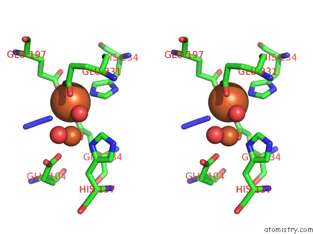
Stereo pair view
|
A full contact list of Iron with other atoms in the Fe binding
site number 3 of Crystal Structure of Toluene 4-Monoxygenase Hydroxylase within 5.0Å range:
probe
|
atom
|
residue
|
distance (Å)
|
B
|
Occ
|
D:Fe507
b:23.0
occ:1.00
|
OE2
|
D:GLU197
|
1.9
|
28.8
|
1.0
|
O
|
D:HOH1349
|
2.0
|
20.6
|
1.0
|
NE2
|
D:HIS234
|
2.2
|
18.7
|
1.0
|
OE2
|
D:GLU231
|
2.2
|
27.3
|
1.0
|
OE2
|
D:GLU134
|
2.2
|
17.9
|
1.0
|
N1
|
D:AZI510
|
2.3
|
33.6
|
1.0
|
CD
|
D:GLU197
|
3.0
|
26.0
|
1.0
|
CD2
|
D:HIS234
|
3.1
|
18.0
|
1.0
|
CD
|
D:GLU231
|
3.1
|
29.1
|
1.0
|
CE1
|
D:HIS234
|
3.1
|
17.8
|
1.0
|
CD
|
D:GLU134
|
3.1
|
16.0
|
1.0
|
FE
|
D:FE508
|
3.2
|
18.9
|
1.0
|
OE1
|
D:GLU134
|
3.4
|
18.9
|
1.0
|
N2
|
D:AZI510
|
3.4
|
42.9
|
1.0
|
OE1
|
D:GLU231
|
3.4
|
33.2
|
1.0
|
O
|
D:HOH1010
|
3.7
|
19.8
|
1.0
|
OE1
|
D:GLU197
|
3.8
|
30.3
|
1.0
|
CG
|
D:GLU197
|
3.9
|
23.2
|
1.0
|
ND1
|
D:HIS234
|
4.3
|
16.4
|
1.0
|
CG
|
D:HIS234
|
4.3
|
19.0
|
1.0
|
O
|
D:HOH657
|
4.4
|
20.0
|
1.0
|
CG
|
D:GLU231
|
4.5
|
22.1
|
1.0
|
CG
|
D:GLU134
|
4.5
|
13.6
|
1.0
|
N3
|
D:AZI510
|
4.5
|
41.6
|
1.0
|
CB
|
D:GLU197
|
4.6
|
19.9
|
1.0
|
ND1
|
D:HIS137
|
4.7
|
15.1
|
1.0
|
CE1
|
D:HIS137
|
4.9
|
15.3
|
1.0
|
OE1
|
D:GLU104
|
4.9
|
20.9
|
1.0
|
|
Iron binding site 4 out
of 4 in 3dhg
Go back to
Iron Binding Sites List in 3dhg
Iron binding site 4 out
of 4 in the Crystal Structure of Toluene 4-Monoxygenase Hydroxylase
Mono view
Stereo pair view
|
A full contact list of Iron with other atoms in the Fe binding
site number 4 of Crystal Structure of Toluene 4-Monoxygenase Hydroxylase within 5.0Å range:
probe
|
atom
|
residue
|
distance (Å)
|
B
|
Occ
|
D:Fe508
b:18.9
occ:1.00
|
OE1
|
D:GLU134
|
2.1
|
18.9
|
1.0
|
OE1
|
D:GLU104
|
2.1
|
20.9
|
1.0
|
O
|
D:HOH1349
|
2.1
|
20.6
|
1.0
|
ND1
|
D:HIS137
|
2.2
|
15.1
|
1.0
|
O
|
D:HOH657
|
2.2
|
20.0
|
1.0
|
N1
|
D:AZI510
|
2.6
|
33.6
|
1.0
|
CD
|
D:GLU104
|
3.1
|
19.2
|
1.0
|
CE1
|
D:HIS137
|
3.1
|
15.3
|
1.0
|
CD
|
D:GLU134
|
3.1
|
16.0
|
1.0
|
FE
|
D:FE507
|
3.2
|
23.0
|
1.0
|
CG
|
D:HIS137
|
3.2
|
16.3
|
1.0
|
N2
|
D:AZI510
|
3.4
|
42.9
|
1.0
|
OE2
|
D:GLU104
|
3.4
|
17.8
|
1.0
|
OE2
|
D:GLU134
|
3.5
|
17.9
|
1.0
|
CB
|
D:HIS137
|
3.6
|
15.0
|
1.0
|
OE1
|
D:GLU231
|
4.2
|
33.2
|
1.0
|
NE2
|
D:HIS137
|
4.3
|
18.1
|
1.0
|
N3
|
D:AZI510
|
4.3
|
41.6
|
1.0
|
CD2
|
D:HIS137
|
4.3
|
17.1
|
1.0
|
CG
|
D:GLU104
|
4.5
|
15.9
|
1.0
|
CG
|
D:GLU134
|
4.5
|
13.6
|
1.0
|
NE2
|
D:HIS234
|
4.5
|
18.7
|
1.0
|
OE2
|
D:GLU231
|
4.6
|
27.3
|
1.0
|
CA
|
D:GLU134
|
4.7
|
14.1
|
1.0
|
CE1
|
D:HIS234
|
4.7
|
17.8
|
1.0
|
CG2
|
D:ILE227
|
4.7
|
17.5
|
1.0
|
CB
|
D:GLU104
|
4.8
|
16.6
|
1.0
|
OE2
|
D:GLU197
|
4.8
|
28.8
|
1.0
|
CD
|
D:GLU231
|
4.9
|
29.1
|
1.0
|
CB
|
D:GLU134
|
4.9
|
14.1
|
1.0
|
O
|
D:ASP133
|
5.0
|
14.4
|
1.0
|
|
Reference:
L.J.Bailey,
J.G.Mccoy,
G.N.Phillips Jr.,
B.G.Fox.
Structural Consequences of Effector Protein Complex Formation in A Diiron Hydroxylase. Proc.Natl.Acad.Sci.Usa V. 105 19194 2008.
ISSN: ISSN 0027-8424
PubMed: 19033467
DOI: 10.1073/PNAS.0807948105
Page generated: Sun Aug 4 09:05:37 2024
|