Iron »
PDB 3dcp-3e0f »
3dhh »
Iron in PDB 3dhh: Crystal Structure of Resting State Toluene 4-Monoxygenase Hydroxylase Complexed with Effector Protein
Protein crystallography data
The structure of Crystal Structure of Resting State Toluene 4-Monoxygenase Hydroxylase Complexed with Effector Protein, PDB code: 3dhh
was solved by
L.J.Bailey,
J.G.Mccoy,
G.N.Phillips Jr.,
B.G.Fox,
with X-Ray Crystallography technique. A brief refinement statistics is given in the table below:
| Resolution Low / High (Å) | 91.29 / 1.94 |
| Space group | C 2 2 21 |
| Cell size a, b, c (Å), α, β, γ (°) | 100.418, 115.613, 182.403, 90.00, 90.00, 90.00 |
| R / Rfree (%) | 15.7 / 20.2 |
Other elements in 3dhh:
The structure of Crystal Structure of Resting State Toluene 4-Monoxygenase Hydroxylase Complexed with Effector Protein also contains other interesting chemical elements:
| Bromine | (Br) | 5 atoms |
| Chlorine | (Cl) | 1 atom |
Iron Binding Sites:
The binding sites of Iron atom in the Crystal Structure of Resting State Toluene 4-Monoxygenase Hydroxylase Complexed with Effector Protein
(pdb code 3dhh). This binding sites where shown within
5.0 Angstroms radius around Iron atom.
In total 2 binding sites of Iron where determined in the Crystal Structure of Resting State Toluene 4-Monoxygenase Hydroxylase Complexed with Effector Protein, PDB code: 3dhh:
Jump to Iron binding site number: 1; 2;
In total 2 binding sites of Iron where determined in the Crystal Structure of Resting State Toluene 4-Monoxygenase Hydroxylase Complexed with Effector Protein, PDB code: 3dhh:
Jump to Iron binding site number: 1; 2;
Iron binding site 1 out of 2 in 3dhh
Go back to
Iron binding site 1 out
of 2 in the Crystal Structure of Resting State Toluene 4-Monoxygenase Hydroxylase Complexed with Effector Protein
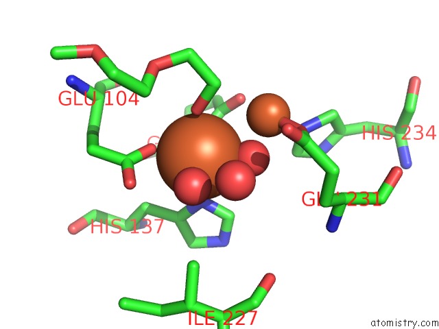
Mono view
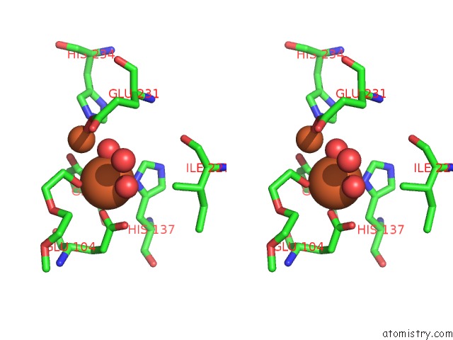
Stereo pair view
Mono view
Stereo pair view
A full contact list of Iron with other atoms in the Fe binding
site number 1 of Crystal Structure of Resting State Toluene 4-Monoxygenase Hydroxylase Complexed with Effector Protein within 5.0Å range:
|
Iron binding site 2 out of 2 in 3dhh
Go back to
Iron binding site 2 out
of 2 in the Crystal Structure of Resting State Toluene 4-Monoxygenase Hydroxylase Complexed with Effector Protein
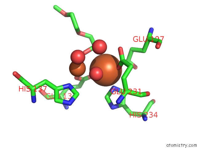
Mono view
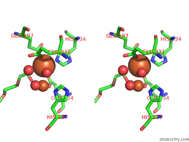
Stereo pair view
Mono view
Stereo pair view
A full contact list of Iron with other atoms in the Fe binding
site number 2 of Crystal Structure of Resting State Toluene 4-Monoxygenase Hydroxylase Complexed with Effector Protein within 5.0Å range:
|
Reference:
L.J.Bailey,
J.G.Mccoy,
G.N.Phillips Jr.,
B.G.Fox.
Structural Consequences of Effector Protein Complex Formation in A Diiron Hydroxylase. Proc.Natl.Acad.Sci.Usa V. 105 19194 2008.
ISSN: ISSN 0027-8424
PubMed: 19033467
DOI: 10.1073/PNAS.0807948105
Page generated: Sun Aug 4 09:05:37 2024
ISSN: ISSN 0027-8424
PubMed: 19033467
DOI: 10.1073/PNAS.0807948105
Last articles
Zn in 9J0NZn in 9J0O
Zn in 9J0P
Zn in 9FJX
Zn in 9EKB
Zn in 9C0F
Zn in 9CAH
Zn in 9CH0
Zn in 9CH3
Zn in 9CH1