Iron »
PDB 3e13-3eai »
3e1p »
Iron in PDB 3e1p: Crystal Structure of E. Coli Bacterioferritin (Bfr) in Which the Ferroxidase Centre Is Inhibited with Zn(II) and High Occupancy Iron Is Bound Within the Cavity.
Protein crystallography data
The structure of Crystal Structure of E. Coli Bacterioferritin (Bfr) in Which the Ferroxidase Centre Is Inhibited with Zn(II) and High Occupancy Iron Is Bound Within the Cavity., PDB code: 3e1p
was solved by
A.Crow,
T.Lawson,
A.Lewin,
G.R.Moore,
N.Le Brun,
with X-Ray Crystallography technique. A brief refinement statistics is given in the table below:
| Resolution Low / High (Å) | 71.25 / 2.40 |
| Space group | P 42 21 2 |
| Cell size a, b, c (Å), α, β, γ (°) | 207.397, 207.397, 142.451, 90.00, 90.00, 90.00 |
| R / Rfree (%) | 24 / 26 |
Other elements in 3e1p:
The structure of Crystal Structure of E. Coli Bacterioferritin (Bfr) in Which the Ferroxidase Centre Is Inhibited with Zn(II) and High Occupancy Iron Is Bound Within the Cavity. also contains other interesting chemical elements:
| Zinc | (Zn) | 24 atoms |
Iron Binding Sites:
Pages:
>>> Page 1 <<< Page 2, Binding sites: 11 - 18;Binding sites:
The binding sites of Iron atom in the Crystal Structure of E. Coli Bacterioferritin (Bfr) in Which the Ferroxidase Centre Is Inhibited with Zn(II) and High Occupancy Iron Is Bound Within the Cavity. (pdb code 3e1p). This binding sites where shown within 5.0 Angstroms radius around Iron atom.In total 18 binding sites of Iron where determined in the Crystal Structure of E. Coli Bacterioferritin (Bfr) in Which the Ferroxidase Centre Is Inhibited with Zn(II) and High Occupancy Iron Is Bound Within the Cavity., PDB code: 3e1p:
Jump to Iron binding site number: 1; 2; 3; 4; 5; 6; 7; 8; 9; 10;
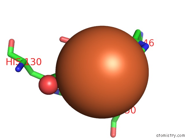
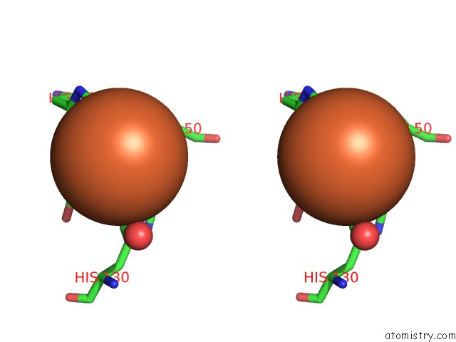
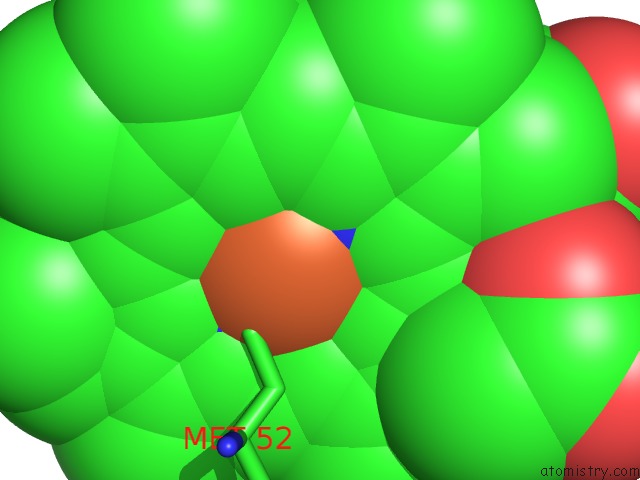
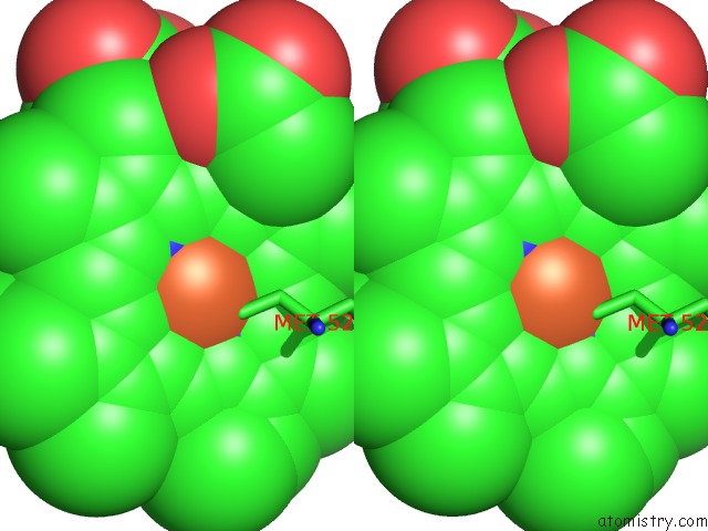
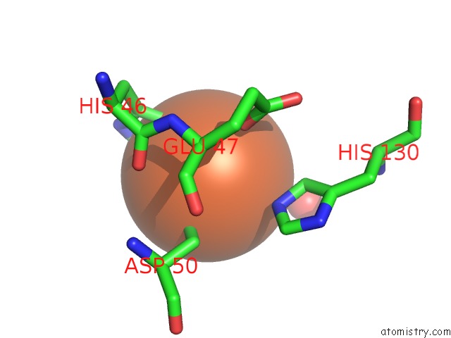
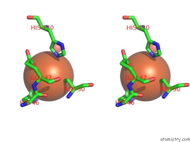
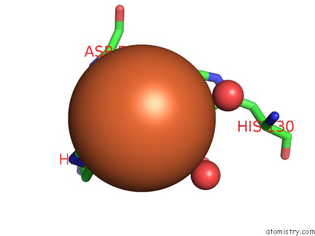
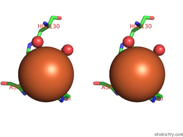
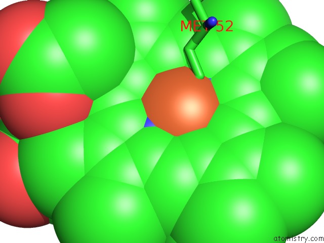
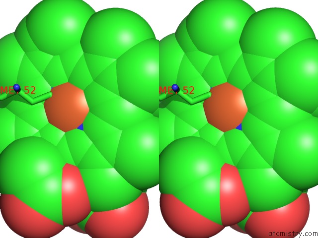
Iron binding site 1 out of 18 in 3e1p
Go back to
Iron binding site 1 out
of 18 in the Crystal Structure of E. Coli Bacterioferritin (Bfr) in Which the Ferroxidase Centre Is Inhibited with Zn(II) and High Occupancy Iron Is Bound Within the Cavity.
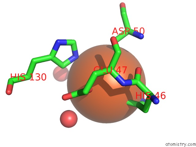
Mono view
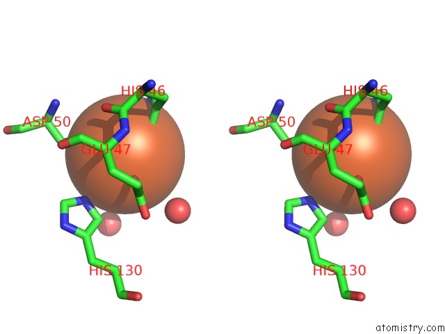
Stereo pair view
Mono view
Stereo pair view
A full contact list of Iron with other atoms in the Fe binding
site number 1 of Crystal Structure of E. Coli Bacterioferritin (Bfr) in Which the Ferroxidase Centre Is Inhibited with Zn(II) and High Occupancy Iron Is Bound Within the Cavity. within 5.0Å range:
|
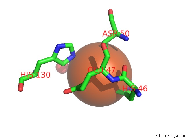
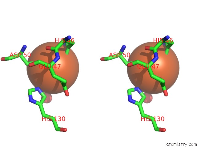
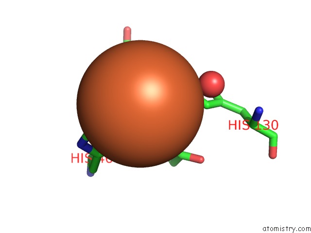
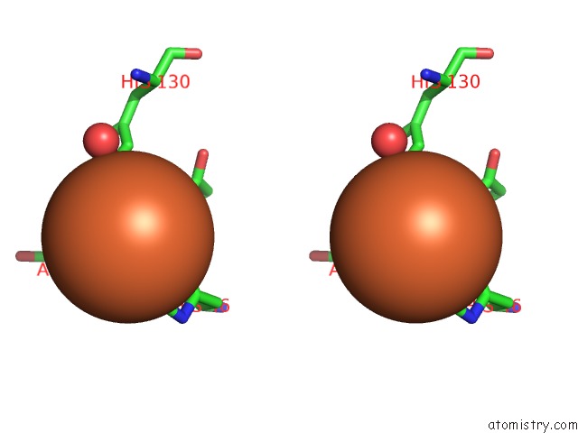
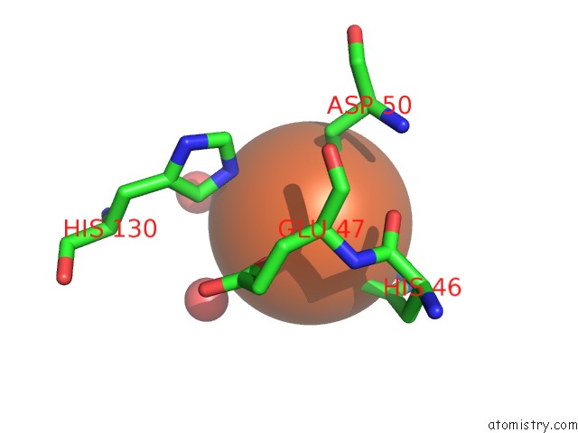
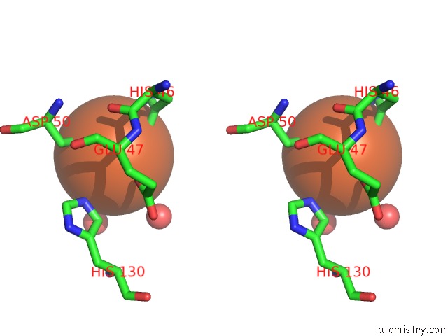
Iron binding site 2 out of 18 in 3e1p
Go back to
Iron binding site 2 out
of 18 in the Crystal Structure of E. Coli Bacterioferritin (Bfr) in Which the Ferroxidase Centre Is Inhibited with Zn(II) and High Occupancy Iron Is Bound Within the Cavity.
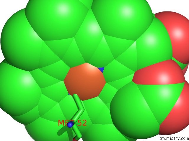
Mono view
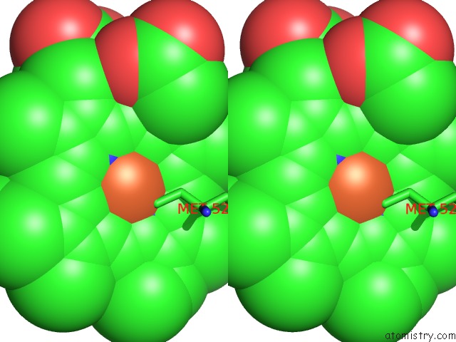
Stereo pair view
Mono view
Stereo pair view
A full contact list of Iron with other atoms in the Fe binding
site number 2 of Crystal Structure of E. Coli Bacterioferritin (Bfr) in Which the Ferroxidase Centre Is Inhibited with Zn(II) and High Occupancy Iron Is Bound Within the Cavity. within 5.0Å range:
|
Iron binding site 3 out of 18 in 3e1p
Go back to
Iron binding site 3 out
of 18 in the Crystal Structure of E. Coli Bacterioferritin (Bfr) in Which the Ferroxidase Centre Is Inhibited with Zn(II) and High Occupancy Iron Is Bound Within the Cavity.
Mono view
Stereo pair view
Mono view
Stereo pair view
A full contact list of Iron with other atoms in the Fe binding
site number 3 of Crystal Structure of E. Coli Bacterioferritin (Bfr) in Which the Ferroxidase Centre Is Inhibited with Zn(II) and High Occupancy Iron Is Bound Within the Cavity. within 5.0Å range:
|
Iron binding site 4 out of 18 in 3e1p
Go back to
Iron binding site 4 out
of 18 in the Crystal Structure of E. Coli Bacterioferritin (Bfr) in Which the Ferroxidase Centre Is Inhibited with Zn(II) and High Occupancy Iron Is Bound Within the Cavity.
Mono view
Stereo pair view
Mono view
Stereo pair view
A full contact list of Iron with other atoms in the Fe binding
site number 4 of Crystal Structure of E. Coli Bacterioferritin (Bfr) in Which the Ferroxidase Centre Is Inhibited with Zn(II) and High Occupancy Iron Is Bound Within the Cavity. within 5.0Å range:
|
Iron binding site 5 out of 18 in 3e1p
Go back to
Iron binding site 5 out
of 18 in the Crystal Structure of E. Coli Bacterioferritin (Bfr) in Which the Ferroxidase Centre Is Inhibited with Zn(II) and High Occupancy Iron Is Bound Within the Cavity.
Mono view
Stereo pair view
Mono view
Stereo pair view
A full contact list of Iron with other atoms in the Fe binding
site number 5 of Crystal Structure of E. Coli Bacterioferritin (Bfr) in Which the Ferroxidase Centre Is Inhibited with Zn(II) and High Occupancy Iron Is Bound Within the Cavity. within 5.0Å range:
|
Iron binding site 6 out of 18 in 3e1p
Go back to
Iron binding site 6 out
of 18 in the Crystal Structure of E. Coli Bacterioferritin (Bfr) in Which the Ferroxidase Centre Is Inhibited with Zn(II) and High Occupancy Iron Is Bound Within the Cavity.
Mono view
Stereo pair view
Mono view
Stereo pair view
A full contact list of Iron with other atoms in the Fe binding
site number 6 of Crystal Structure of E. Coli Bacterioferritin (Bfr) in Which the Ferroxidase Centre Is Inhibited with Zn(II) and High Occupancy Iron Is Bound Within the Cavity. within 5.0Å range:
|
Iron binding site 7 out of 18 in 3e1p
Go back to
Iron binding site 7 out
of 18 in the Crystal Structure of E. Coli Bacterioferritin (Bfr) in Which the Ferroxidase Centre Is Inhibited with Zn(II) and High Occupancy Iron Is Bound Within the Cavity.
Mono view
Stereo pair view
Mono view
Stereo pair view
A full contact list of Iron with other atoms in the Fe binding
site number 7 of Crystal Structure of E. Coli Bacterioferritin (Bfr) in Which the Ferroxidase Centre Is Inhibited with Zn(II) and High Occupancy Iron Is Bound Within the Cavity. within 5.0Å range:
|
Iron binding site 8 out of 18 in 3e1p
Go back to
Iron binding site 8 out
of 18 in the Crystal Structure of E. Coli Bacterioferritin (Bfr) in Which the Ferroxidase Centre Is Inhibited with Zn(II) and High Occupancy Iron Is Bound Within the Cavity.
Mono view
Stereo pair view
Mono view
Stereo pair view
A full contact list of Iron with other atoms in the Fe binding
site number 8 of Crystal Structure of E. Coli Bacterioferritin (Bfr) in Which the Ferroxidase Centre Is Inhibited with Zn(II) and High Occupancy Iron Is Bound Within the Cavity. within 5.0Å range:
|
Iron binding site 9 out of 18 in 3e1p
Go back to
Iron binding site 9 out
of 18 in the Crystal Structure of E. Coli Bacterioferritin (Bfr) in Which the Ferroxidase Centre Is Inhibited with Zn(II) and High Occupancy Iron Is Bound Within the Cavity.
Mono view
Stereo pair view
Mono view
Stereo pair view
A full contact list of Iron with other atoms in the Fe binding
site number 9 of Crystal Structure of E. Coli Bacterioferritin (Bfr) in Which the Ferroxidase Centre Is Inhibited with Zn(II) and High Occupancy Iron Is Bound Within the Cavity. within 5.0Å range:
|
Iron binding site 10 out of 18 in 3e1p
Go back to
Iron binding site 10 out
of 18 in the Crystal Structure of E. Coli Bacterioferritin (Bfr) in Which the Ferroxidase Centre Is Inhibited with Zn(II) and High Occupancy Iron Is Bound Within the Cavity.
Mono view
Stereo pair view
Mono view
Stereo pair view
A full contact list of Iron with other atoms in the Fe binding
site number 10 of Crystal Structure of E. Coli Bacterioferritin (Bfr) in Which the Ferroxidase Centre Is Inhibited with Zn(II) and High Occupancy Iron Is Bound Within the Cavity. within 5.0Å range:
|
Reference:
A.Crow,
T.L.Lawson,
A.Lewin,
G.R.Moore,
N.E.Le Brun.
Structural Basis For Iron Mineralization By Bacterioferritin J.Am.Chem.Soc. V. 131 6808 2009.
ISSN: ISSN 0002-7863
PubMed: 19391621
DOI: 10.1021/JA8093444
Page generated: Sun Aug 4 09:17:50 2024
ISSN: ISSN 0002-7863
PubMed: 19391621
DOI: 10.1021/JA8093444
Last articles
Cl in 3PUSCl in 3PUG
Cl in 3PVC
Cl in 3PUP
Cl in 3PPC
Cl in 3PU8
Cl in 3PUA
Cl in 3PSQ
Cl in 3PSU
Cl in 3PS9