Iron »
PDB 3e13-3eai »
3e4y »
Iron in PDB 3e4y: Crystal Structure of A 33KDA Catalase-Related Protein From Mycobacterium Avium Subsp. Paratuberculosis. I2(1)2(1)2(1) Crystal Form
Protein crystallography data
The structure of Crystal Structure of A 33KDA Catalase-Related Protein From Mycobacterium Avium Subsp. Paratuberculosis. I2(1)2(1)2(1) Crystal Form, PDB code: 3e4y
was solved by
S.Pakhomova,
M.E.Newcomer,
with X-Ray Crystallography technique. A brief refinement statistics is given in the table below:
| Resolution Low / High (Å) | 43.20 / 2.60 |
| Space group | I 21 21 21 |
| Cell size a, b, c (Å), α, β, γ (°) | 69.604, 92.680, 165.499, 90.00, 90.00, 90.00 |
| R / Rfree (%) | 20.2 / 25.7 |
Iron Binding Sites:
The binding sites of Iron atom in the Crystal Structure of A 33KDA Catalase-Related Protein From Mycobacterium Avium Subsp. Paratuberculosis. I2(1)2(1)2(1) Crystal Form
(pdb code 3e4y). This binding sites where shown within
5.0 Angstroms radius around Iron atom.
In total only one binding site of Iron was determined in the Crystal Structure of A 33KDA Catalase-Related Protein From Mycobacterium Avium Subsp. Paratuberculosis. I2(1)2(1)2(1) Crystal Form, PDB code: 3e4y:
In total only one binding site of Iron was determined in the Crystal Structure of A 33KDA Catalase-Related Protein From Mycobacterium Avium Subsp. Paratuberculosis. I2(1)2(1)2(1) Crystal Form, PDB code: 3e4y:
Iron binding site 1 out of 1 in 3e4y
Go back to
Iron binding site 1 out
of 1 in the Crystal Structure of A 33KDA Catalase-Related Protein From Mycobacterium Avium Subsp. Paratuberculosis. I2(1)2(1)2(1) Crystal Form
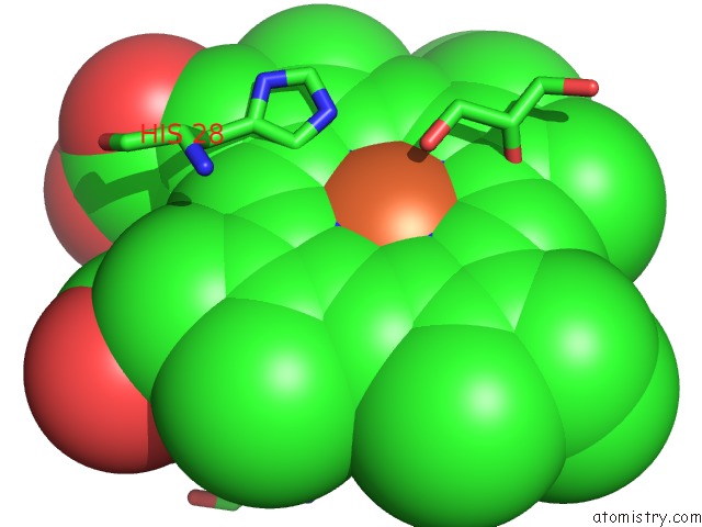
Mono view
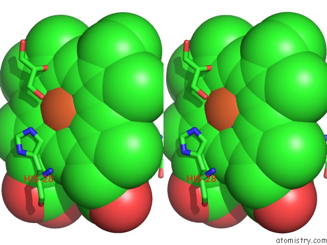
Stereo pair view
Mono view
Stereo pair view
A full contact list of Iron with other atoms in the Fe binding
site number 1 of Crystal Structure of A 33KDA Catalase-Related Protein From Mycobacterium Avium Subsp. Paratuberculosis. I2(1)2(1)2(1) Crystal Form within 5.0Å range:
|
Reference:
S.Pakhomova,
B.Gao,
W.E.Boeglin,
A.R.Brash,
M.E.Newcomer.
The Structure and Peroxidase Activity of A 33-kDa Catalase-Related Protein From Mycobacterium Avium Ssp. Paratuberculosis. Protein Sci. V. 18 2559 2009.
ISSN: ISSN 0961-8368
PubMed: 19827095
DOI: 10.1002/PRO.265
Page generated: Sun Aug 4 09:21:16 2024
ISSN: ISSN 0961-8368
PubMed: 19827095
DOI: 10.1002/PRO.265
Last articles
Zn in 9MJ5Zn in 9HNW
Zn in 9G0L
Zn in 9FNE
Zn in 9DZN
Zn in 9E0I
Zn in 9D32
Zn in 9DAK
Zn in 8ZXC
Zn in 8ZUF