Iron »
PDB 3e13-3eai »
3e5o »
Iron in PDB 3e5o: Carbonmonoxy Sperm Whale Myoglobin at 140 K: Laser Off
Protein crystallography data
The structure of Carbonmonoxy Sperm Whale Myoglobin at 140 K: Laser Off, PDB code: 3e5o
was solved by
A.Tomita,
T.Sato,
K.Ichiyanagi,
S.Nozawa,
H.Ichikawa,
M.Chollet,
F.Kawai,
S.-Y.Park,
S.Koshihara,
S.Adachi,
with X-Ray Crystallography technique. A brief refinement statistics is given in the table below:
| Resolution Low / High (Å) | 17.01 / 1.21 |
| Space group | P 1 21 1 |
| Cell size a, b, c (Å), α, β, γ (°) | 34.343, 30.613, 63.785, 90.00, 105.86, 90.00 |
| R / Rfree (%) | 15.6 / 20.9 |
Iron Binding Sites:
The binding sites of Iron atom in the Carbonmonoxy Sperm Whale Myoglobin at 140 K: Laser Off
(pdb code 3e5o). This binding sites where shown within
5.0 Angstroms radius around Iron atom.
In total only one binding site of Iron was determined in the Carbonmonoxy Sperm Whale Myoglobin at 140 K: Laser Off, PDB code: 3e5o:
In total only one binding site of Iron was determined in the Carbonmonoxy Sperm Whale Myoglobin at 140 K: Laser Off, PDB code: 3e5o:
Iron binding site 1 out of 1 in 3e5o
Go back to
Iron binding site 1 out
of 1 in the Carbonmonoxy Sperm Whale Myoglobin at 140 K: Laser Off
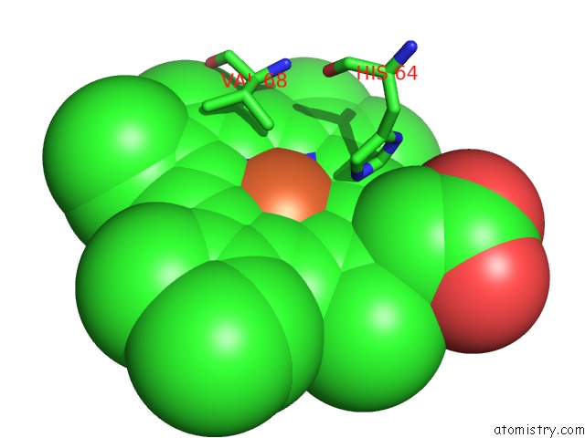
Mono view
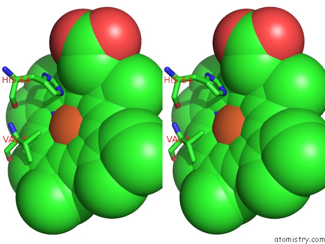
Stereo pair view
Mono view
Stereo pair view
|
|
A full contact list of Iron with other atoms in the Fe binding
site number 1 of Carbonmonoxy Sperm Whale Myoglobin at 140 K: Laser Off within 5.0Å range:
|
Reference:
A.Tomita,
T.Sato,
K.Ichiyanagi,
S.Nozawa,
H.Ichikawa,
M.Chollet,
F.Kawai,
S.-Y.Park,
T.Tsuduki,
T.Yamato,
S.Koshihara,
S.Adachi.
Visualizing Breathing Motion of Internal Cavities in Concert with Ligand Migration in Myoglobin. Proc.Natl.Acad.Sci.Usa V. 106 2612 2009.
ISSN: ISSN 0027-8424
PubMed: 19204297
DOI: 10.1073/PNAS.0807774106
Page generated: Tue Aug 5 00:52:08 2025
ISSN: ISSN 0027-8424
PubMed: 19204297
DOI: 10.1073/PNAS.0807774106
Last articles
Zn in 7KD2Zn in 7KDU
Zn in 7KD0
Zn in 7KCY
Zn in 7KCX
Zn in 7KCB
Zn in 7KCQ
Zn in 7KCW
Zn in 7KCV
Zn in 7KC9