Iron »
PDB 3ebd-3esf »
3ecj »
Iron in PDB 3ecj: Structure of E323L Mutant of Homoprotocatechuate 2,3-Dioxygenase From Brevibacterium Fuscum at 1.65A Resolution
Enzymatic activity of Structure of E323L Mutant of Homoprotocatechuate 2,3-Dioxygenase From Brevibacterium Fuscum at 1.65A Resolution
All present enzymatic activity of Structure of E323L Mutant of Homoprotocatechuate 2,3-Dioxygenase From Brevibacterium Fuscum at 1.65A Resolution:
1.13.11.15;
1.13.11.15;
Protein crystallography data
The structure of Structure of E323L Mutant of Homoprotocatechuate 2,3-Dioxygenase From Brevibacterium Fuscum at 1.65A Resolution, PDB code: 3ecj
was solved by
E.G.Kovaleva,
J.D.Lipscomb,
with X-Ray Crystallography technique. A brief refinement statistics is given in the table below:
| Resolution Low / High (Å) | 23.46 / 1.65 |
| Space group | P 21 21 2 |
| Cell size a, b, c (Å), α, β, γ (°) | 110.711, 163.403, 101.584, 90.00, 90.00, 90.00 |
| R / Rfree (%) | 16.9 / 19.4 |
Other elements in 3ecj:
The structure of Structure of E323L Mutant of Homoprotocatechuate 2,3-Dioxygenase From Brevibacterium Fuscum at 1.65A Resolution also contains other interesting chemical elements:
| Chlorine | (Cl) | 4 atoms |
| Calcium | (Ca) | 1 atom |
Iron Binding Sites:
The binding sites of Iron atom in the Structure of E323L Mutant of Homoprotocatechuate 2,3-Dioxygenase From Brevibacterium Fuscum at 1.65A Resolution
(pdb code 3ecj). This binding sites where shown within
5.0 Angstroms radius around Iron atom.
In total 4 binding sites of Iron where determined in the Structure of E323L Mutant of Homoprotocatechuate 2,3-Dioxygenase From Brevibacterium Fuscum at 1.65A Resolution, PDB code: 3ecj:
Jump to Iron binding site number: 1; 2; 3; 4;
In total 4 binding sites of Iron where determined in the Structure of E323L Mutant of Homoprotocatechuate 2,3-Dioxygenase From Brevibacterium Fuscum at 1.65A Resolution, PDB code: 3ecj:
Jump to Iron binding site number: 1; 2; 3; 4;
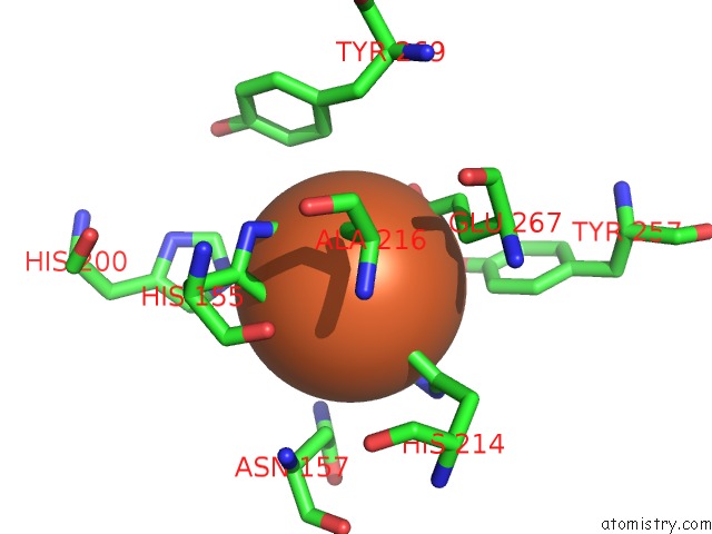
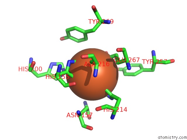
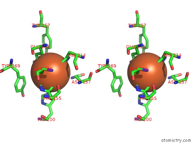
Iron binding site 1 out of 4 in 3ecj
Go back to
Iron binding site 1 out
of 4 in the Structure of E323L Mutant of Homoprotocatechuate 2,3-Dioxygenase From Brevibacterium Fuscum at 1.65A Resolution
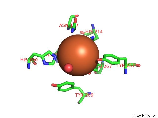
Mono view
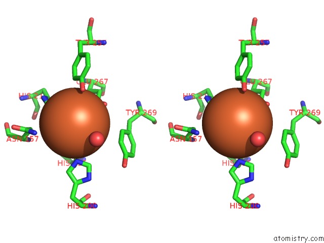
Stereo pair view
Mono view
Stereo pair view
A full contact list of Iron with other atoms in the Fe binding
site number 1 of Structure of E323L Mutant of Homoprotocatechuate 2,3-Dioxygenase From Brevibacterium Fuscum at 1.65A Resolution within 5.0Å range:
|
Iron binding site 2 out of 4 in 3ecj
Go back to
Iron binding site 2 out
of 4 in the Structure of E323L Mutant of Homoprotocatechuate 2,3-Dioxygenase From Brevibacterium Fuscum at 1.65A Resolution
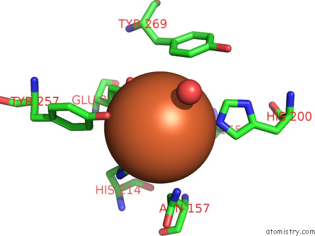
Mono view
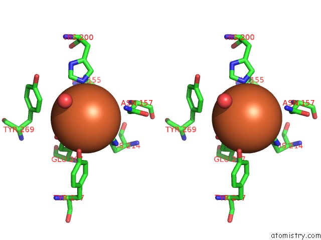
Stereo pair view
Mono view
Stereo pair view
A full contact list of Iron with other atoms in the Fe binding
site number 2 of Structure of E323L Mutant of Homoprotocatechuate 2,3-Dioxygenase From Brevibacterium Fuscum at 1.65A Resolution within 5.0Å range:
|
Iron binding site 3 out of 4 in 3ecj
Go back to
Iron binding site 3 out
of 4 in the Structure of E323L Mutant of Homoprotocatechuate 2,3-Dioxygenase From Brevibacterium Fuscum at 1.65A Resolution
Mono view

Stereo pair view
Mono view
Stereo pair view
A full contact list of Iron with other atoms in the Fe binding
site number 3 of Structure of E323L Mutant of Homoprotocatechuate 2,3-Dioxygenase From Brevibacterium Fuscum at 1.65A Resolution within 5.0Å range:
|
Iron binding site 4 out of 4 in 3ecj
Go back to
Iron binding site 4 out
of 4 in the Structure of E323L Mutant of Homoprotocatechuate 2,3-Dioxygenase From Brevibacterium Fuscum at 1.65A Resolution
Mono view
Stereo pair view
Mono view
Stereo pair view
A full contact list of Iron with other atoms in the Fe binding
site number 4 of Structure of E323L Mutant of Homoprotocatechuate 2,3-Dioxygenase From Brevibacterium Fuscum at 1.65A Resolution within 5.0Å range:
|
Reference:
E.G.Kovaleva,
J.D.Lipscomb.
Intermediate in the O-O Bond Cleavage Reaction of An Extradiol Dioxygenase. Biochemistry V. 47 11168 2008.
ISSN: ISSN 0006-2960
PubMed: 18826259
DOI: 10.1021/BI801459Q
Page generated: Sun Aug 4 09:42:04 2024
ISSN: ISSN 0006-2960
PubMed: 18826259
DOI: 10.1021/BI801459Q
Last articles
Zn in 9J0NZn in 9J0O
Zn in 9J0P
Zn in 9FJX
Zn in 9EKB
Zn in 9C0F
Zn in 9CAH
Zn in 9CH0
Zn in 9CH3
Zn in 9CH1