Iron »
PDB 3ebd-3esf »
3egm »
Iron in PDB 3egm: Structural Basis of Iron Transport Gating in Helicobacter Pylori Ferritin
Enzymatic activity of Structural Basis of Iron Transport Gating in Helicobacter Pylori Ferritin
All present enzymatic activity of Structural Basis of Iron Transport Gating in Helicobacter Pylori Ferritin:
1.16.3.1;
1.16.3.1;
Protein crystallography data
The structure of Structural Basis of Iron Transport Gating in Helicobacter Pylori Ferritin, PDB code: 3egm
was solved by
K.H.Kim,
K.J.Cho,
H.J.Shin,
J.H.Lee,
with X-Ray Crystallography technique. A brief refinement statistics is given in the table below:
| Resolution Low / High (Å) | 29.95 / 2.10 |
| Space group | I 4 |
| Cell size a, b, c (Å), α, β, γ (°) | 128.528, 128.528, 165.380, 90.00, 90.00, 90.00 |
| R / Rfree (%) | 16.2 / 20.4 |
Iron Binding Sites:
Pages:
>>> Page 1 <<< Page 2, Binding sites: 11 - 15;Binding sites:
The binding sites of Iron atom in the Structural Basis of Iron Transport Gating in Helicobacter Pylori Ferritin (pdb code 3egm). This binding sites where shown within 5.0 Angstroms radius around Iron atom.In total 15 binding sites of Iron where determined in the Structural Basis of Iron Transport Gating in Helicobacter Pylori Ferritin, PDB code: 3egm:
Jump to Iron binding site number: 1; 2; 3; 4; 5; 6; 7; 8; 9; 10;
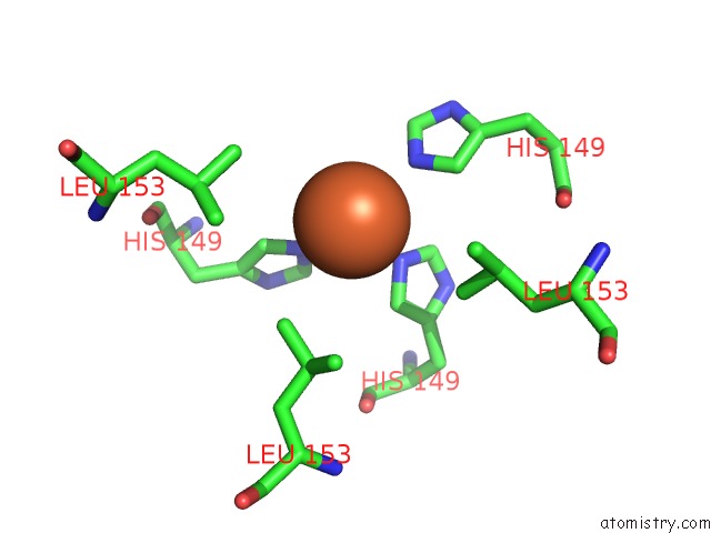
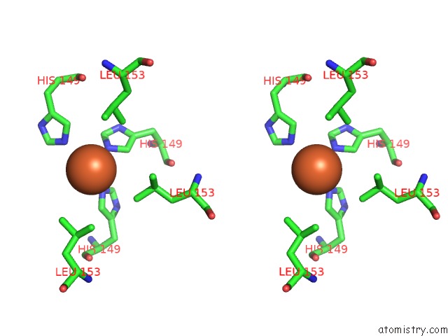
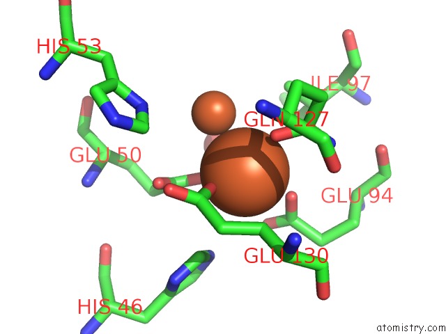
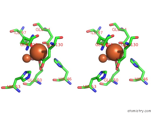
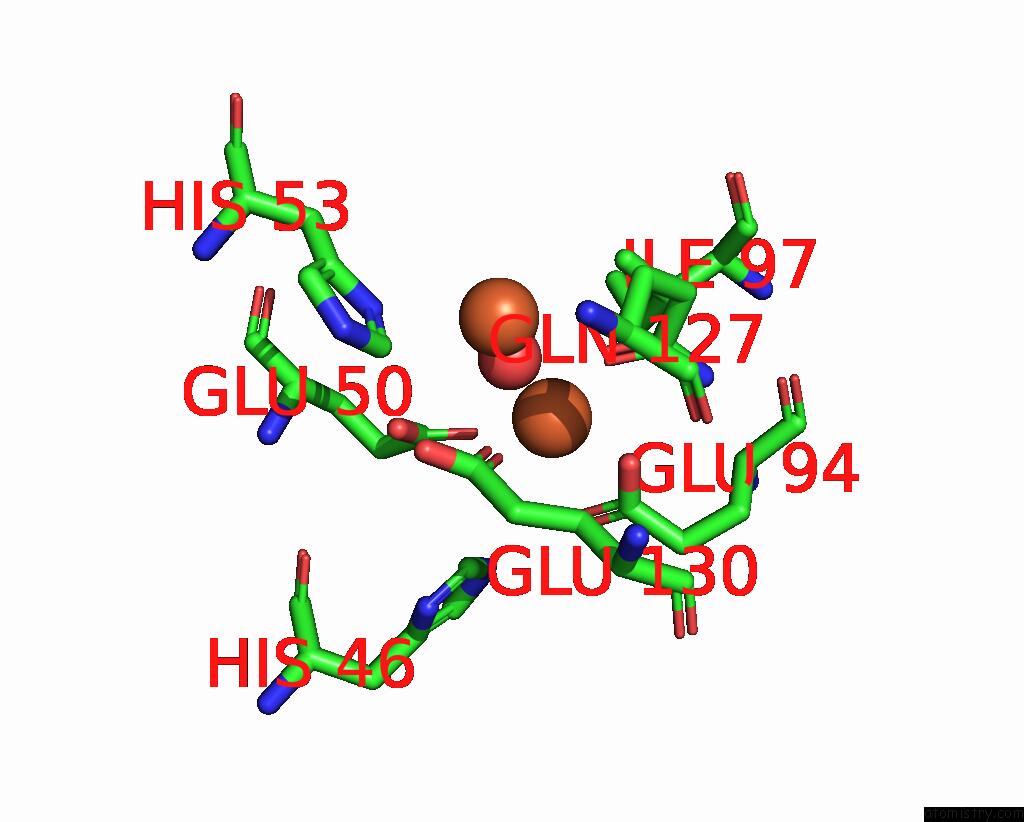
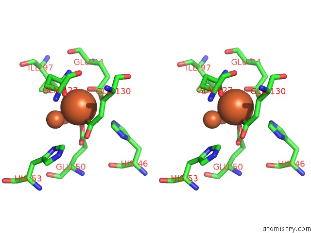
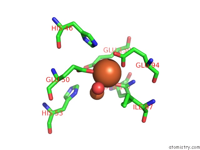
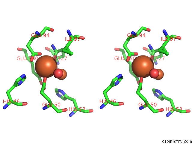
Iron binding site 1 out of 15 in 3egm
Go back to
Iron binding site 1 out
of 15 in the Structural Basis of Iron Transport Gating in Helicobacter Pylori Ferritin
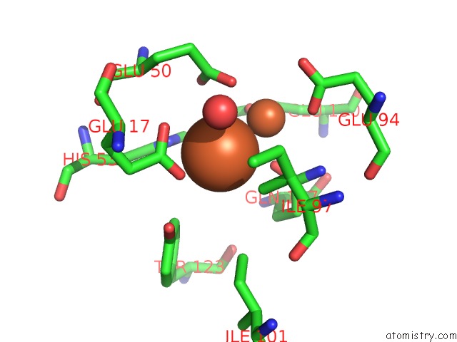
Mono view
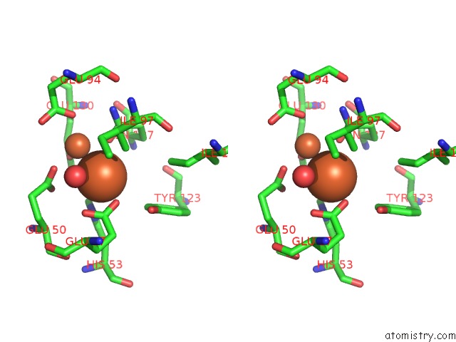
Stereo pair view
Mono view
Stereo pair view
A full contact list of Iron with other atoms in the Fe binding
site number 1 of Structural Basis of Iron Transport Gating in Helicobacter Pylori Ferritin within 5.0Å range:
|
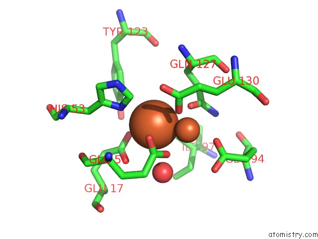
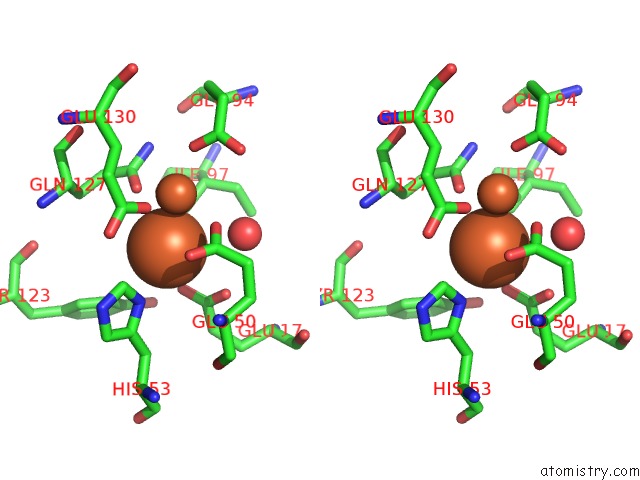
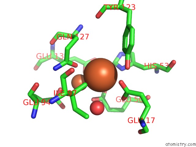
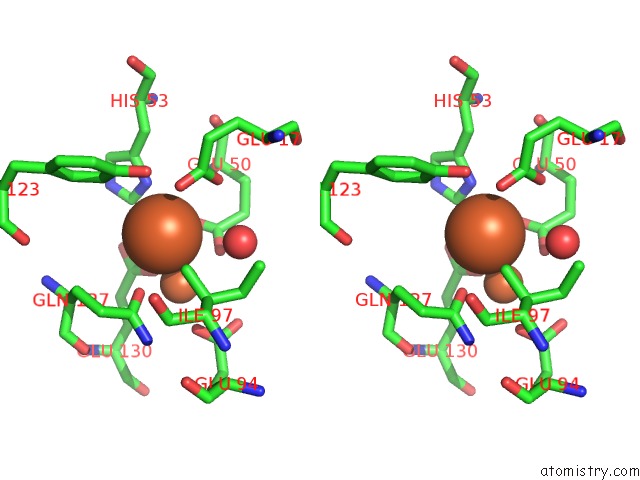
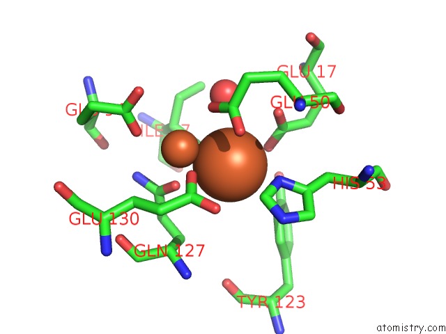
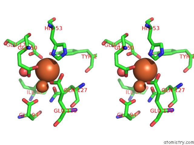
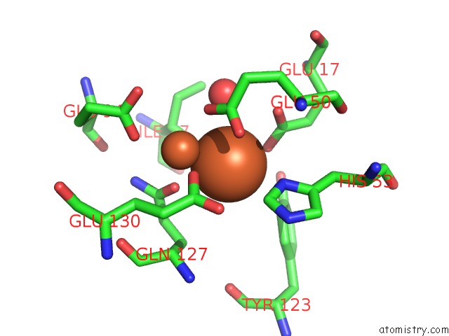
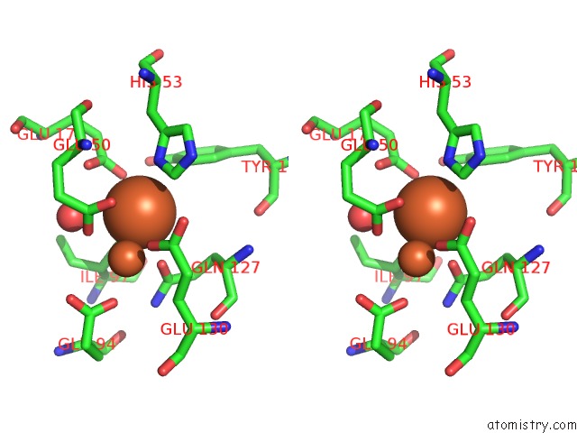
Iron binding site 2 out of 15 in 3egm
Go back to
Iron binding site 2 out
of 15 in the Structural Basis of Iron Transport Gating in Helicobacter Pylori Ferritin
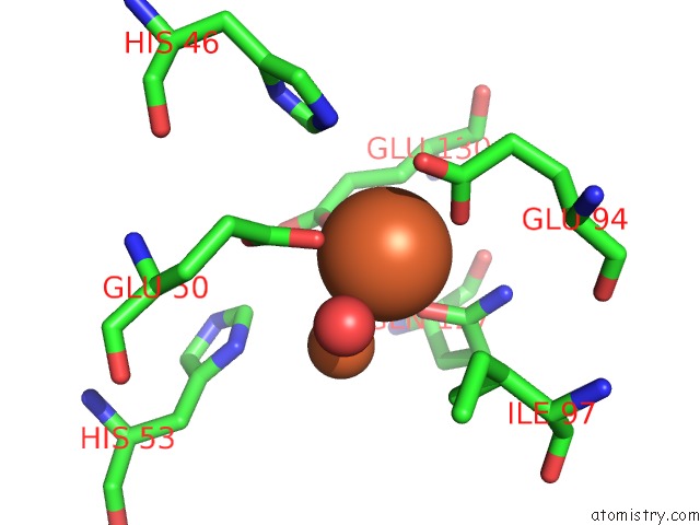
Mono view
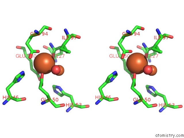
Stereo pair view
Mono view
Stereo pair view
A full contact list of Iron with other atoms in the Fe binding
site number 2 of Structural Basis of Iron Transport Gating in Helicobacter Pylori Ferritin within 5.0Å range:
|
Iron binding site 3 out of 15 in 3egm
Go back to
Iron binding site 3 out
of 15 in the Structural Basis of Iron Transport Gating in Helicobacter Pylori Ferritin
Mono view
Stereo pair view
Mono view
Stereo pair view
A full contact list of Iron with other atoms in the Fe binding
site number 3 of Structural Basis of Iron Transport Gating in Helicobacter Pylori Ferritin within 5.0Å range:
|
Iron binding site 4 out of 15 in 3egm
Go back to
Iron binding site 4 out
of 15 in the Structural Basis of Iron Transport Gating in Helicobacter Pylori Ferritin
Mono view
Stereo pair view
Mono view
Stereo pair view
A full contact list of Iron with other atoms in the Fe binding
site number 4 of Structural Basis of Iron Transport Gating in Helicobacter Pylori Ferritin within 5.0Å range:
|
Iron binding site 5 out of 15 in 3egm
Go back to
Iron binding site 5 out
of 15 in the Structural Basis of Iron Transport Gating in Helicobacter Pylori Ferritin
Mono view
Stereo pair view
Mono view
Stereo pair view
A full contact list of Iron with other atoms in the Fe binding
site number 5 of Structural Basis of Iron Transport Gating in Helicobacter Pylori Ferritin within 5.0Å range:
|
Iron binding site 6 out of 15 in 3egm
Go back to
Iron binding site 6 out
of 15 in the Structural Basis of Iron Transport Gating in Helicobacter Pylori Ferritin
Mono view
Stereo pair view
Mono view
Stereo pair view
A full contact list of Iron with other atoms in the Fe binding
site number 6 of Structural Basis of Iron Transport Gating in Helicobacter Pylori Ferritin within 5.0Å range:
|
Iron binding site 7 out of 15 in 3egm
Go back to
Iron binding site 7 out
of 15 in the Structural Basis of Iron Transport Gating in Helicobacter Pylori Ferritin
Mono view
Stereo pair view
Mono view
Stereo pair view
A full contact list of Iron with other atoms in the Fe binding
site number 7 of Structural Basis of Iron Transport Gating in Helicobacter Pylori Ferritin within 5.0Å range:
|
Iron binding site 8 out of 15 in 3egm
Go back to
Iron binding site 8 out
of 15 in the Structural Basis of Iron Transport Gating in Helicobacter Pylori Ferritin
Mono view
Stereo pair view
Mono view
Stereo pair view
A full contact list of Iron with other atoms in the Fe binding
site number 8 of Structural Basis of Iron Transport Gating in Helicobacter Pylori Ferritin within 5.0Å range:
|
Iron binding site 9 out of 15 in 3egm
Go back to
Iron binding site 9 out
of 15 in the Structural Basis of Iron Transport Gating in Helicobacter Pylori Ferritin
Mono view
Stereo pair view
Mono view
Stereo pair view
A full contact list of Iron with other atoms in the Fe binding
site number 9 of Structural Basis of Iron Transport Gating in Helicobacter Pylori Ferritin within 5.0Å range:
|
Iron binding site 10 out of 15 in 3egm
Go back to
Iron binding site 10 out
of 15 in the Structural Basis of Iron Transport Gating in Helicobacter Pylori Ferritin
Mono view
Stereo pair view
Mono view
Stereo pair view
A full contact list of Iron with other atoms in the Fe binding
site number 10 of Structural Basis of Iron Transport Gating in Helicobacter Pylori Ferritin within 5.0Å range:
|
Reference:
K.J.Cho,
H.J.Shin,
J.H.Lee,
K.J.Kim,
S.S.Park,
Y.Lee,
C.Lee,
S.S.Park,
K.H.Kim.
The Crystal Structure of Ferritin From Helicobacter Pylori Reveals Unusual Conformational Changes For Iron Uptake. J.Mol.Biol. V. 390 83 2009.
ISSN: ISSN 0022-2836
PubMed: 19427319
DOI: 10.1016/J.JMB.2009.04.078
Page generated: Sun Aug 4 09:43:20 2024
ISSN: ISSN 0022-2836
PubMed: 19427319
DOI: 10.1016/J.JMB.2009.04.078
Last articles
Zn in 9MJ5Zn in 9HNW
Zn in 9G0L
Zn in 9FNE
Zn in 9DZN
Zn in 9E0I
Zn in 9D32
Zn in 9DAK
Zn in 8ZXC
Zn in 8ZUF