Iron »
PDB 3ebd-3esf »
3emm »
Iron in PDB 3emm: X-Ray Structure of Protein From Arabidopsis Thaliana AT1G79260 with Bound Heme
Protein crystallography data
The structure of X-Ray Structure of Protein From Arabidopsis Thaliana AT1G79260 with Bound Heme, PDB code: 3emm
was solved by
C.M.Bianchetti,
C.A.Bingman,
G.E.Wesenberg,
G.N.Phillips Jr.,
Center Foreukaryotic Structural Genomics (Cesg),
with X-Ray Crystallography technique. A brief refinement statistics is given in the table below:
| Resolution Low / High (Å) | 47.84 / 1.36 |
| Space group | P 21 21 2 |
| Cell size a, b, c (Å), α, β, γ (°) | 59.749, 79.732, 36.971, 90.00, 90.00, 90.00 |
| R / Rfree (%) | 17 / 19.8 |
Iron Binding Sites:
The binding sites of Iron atom in the X-Ray Structure of Protein From Arabidopsis Thaliana AT1G79260 with Bound Heme
(pdb code 3emm). This binding sites where shown within
5.0 Angstroms radius around Iron atom.
In total only one binding site of Iron was determined in the X-Ray Structure of Protein From Arabidopsis Thaliana AT1G79260 with Bound Heme, PDB code: 3emm:
In total only one binding site of Iron was determined in the X-Ray Structure of Protein From Arabidopsis Thaliana AT1G79260 with Bound Heme, PDB code: 3emm:
Iron binding site 1 out of 1 in 3emm
Go back to
Iron binding site 1 out
of 1 in the X-Ray Structure of Protein From Arabidopsis Thaliana AT1G79260 with Bound Heme
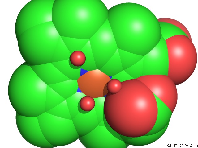
Mono view
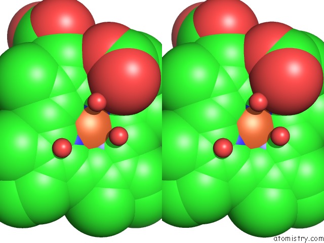
Stereo pair view
Mono view
Stereo pair view
A full contact list of Iron with other atoms in the Fe binding
site number 1 of X-Ray Structure of Protein From Arabidopsis Thaliana AT1G79260 with Bound Heme within 5.0Å range:
|
Reference:
C.M.Bianchetti,
G.C.Blouin,
E.Bitto,
J.S.Olson,
G.N.Phillips.
The Structure and No Binding Properties of the Nitrophorin-Like Heme-Binding Protein From Arabidopsis Thaliana Gene Locus AT1G79260.1. Proteins V. 78 917 2010.
ISSN: ISSN 0887-3585
PubMed: 19938152
DOI: 10.1002/PROT.22617
Page generated: Sun Aug 4 09:49:20 2024
ISSN: ISSN 0887-3585
PubMed: 19938152
DOI: 10.1002/PROT.22617
Last articles
Cl in 2Y2ICl in 2Y1X
Cl in 2Y1G
Cl in 2Y1F
Cl in 2Y05
Cl in 2Y1K
Cl in 2XZR
Cl in 2Y08
Cl in 2Y1D
Cl in 2XZ5