Iron »
PDB 3ebd-3esf »
3en1 »
Iron in PDB 3en1: Crystal Structure of Toluene 2,3-Dioxygenase
Enzymatic activity of Crystal Structure of Toluene 2,3-Dioxygenase
All present enzymatic activity of Crystal Structure of Toluene 2,3-Dioxygenase:
1.14.12.11;
1.14.12.11;
Protein crystallography data
The structure of Crystal Structure of Toluene 2,3-Dioxygenase, PDB code: 3en1
was solved by
R.Friemann,
K.Lee,
E.N.Brown,
D.T.Gibson,
H.Eklund,
S.Ramaswamy,
with X-Ray Crystallography technique. A brief refinement statistics is given in the table below:
| Resolution Low / High (Å) | 20.00 / 3.20 |
| Space group | P 43 3 2 |
| Cell size a, b, c (Å), α, β, γ (°) | 234.531, 234.531, 234.531, 90.00, 90.00, 90.00 |
| R / Rfree (%) | 18.5 / 20.8 |
Iron Binding Sites:
The binding sites of Iron atom in the Crystal Structure of Toluene 2,3-Dioxygenase
(pdb code 3en1). This binding sites where shown within
5.0 Angstroms radius around Iron atom.
In total 4 binding sites of Iron where determined in the Crystal Structure of Toluene 2,3-Dioxygenase, PDB code: 3en1:
Jump to Iron binding site number: 1; 2; 3; 4;
In total 4 binding sites of Iron where determined in the Crystal Structure of Toluene 2,3-Dioxygenase, PDB code: 3en1:
Jump to Iron binding site number: 1; 2; 3; 4;
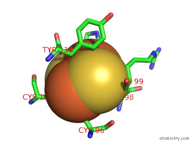
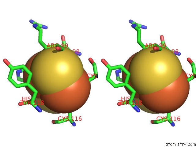
Iron binding site 1 out of 4 in 3en1
Go back to
Iron binding site 1 out
of 4 in the Crystal Structure of Toluene 2,3-Dioxygenase
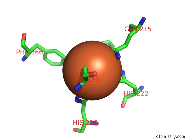
Mono view
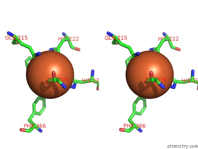
Stereo pair view
Mono view
Stereo pair view
A full contact list of Iron with other atoms in the Fe binding
site number 1 of Crystal Structure of Toluene 2,3-Dioxygenase within 5.0Å range:
|
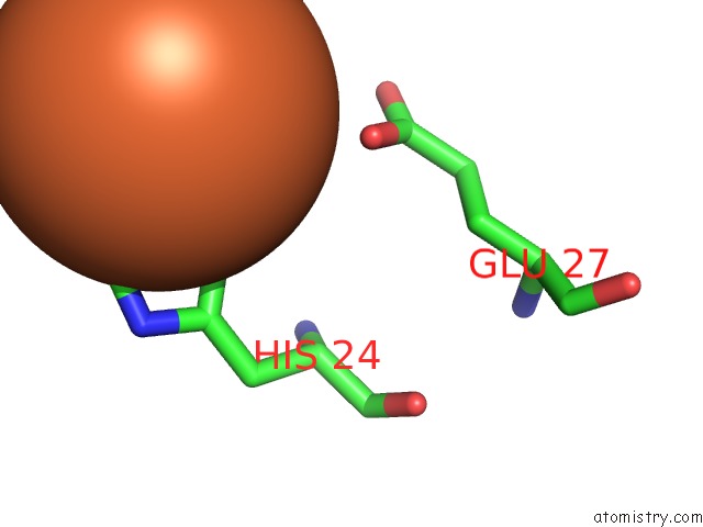
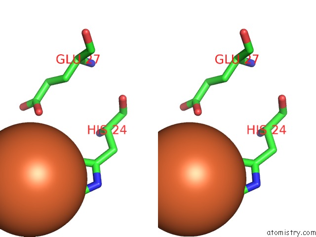
Iron binding site 2 out of 4 in 3en1
Go back to
Iron binding site 2 out
of 4 in the Crystal Structure of Toluene 2,3-Dioxygenase
Mono view
Stereo pair view
Mono view
Stereo pair view
A full contact list of Iron with other atoms in the Fe binding
site number 2 of Crystal Structure of Toluene 2,3-Dioxygenase within 5.0Å range:
|
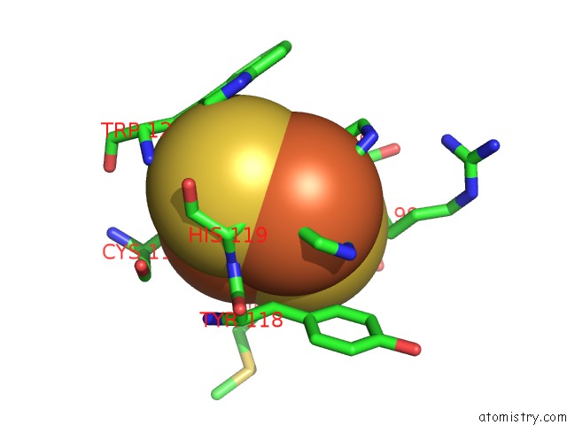
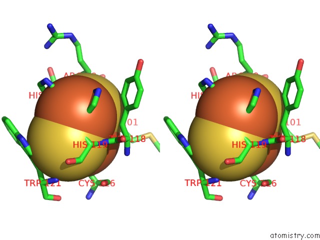
Iron binding site 3 out of 4 in 3en1
Go back to
Iron binding site 3 out
of 4 in the Crystal Structure of Toluene 2,3-Dioxygenase
Mono view
Stereo pair view
Mono view
Stereo pair view
A full contact list of Iron with other atoms in the Fe binding
site number 3 of Crystal Structure of Toluene 2,3-Dioxygenase within 5.0Å range:
|
Iron binding site 4 out of 4 in 3en1
Go back to
Iron binding site 4 out
of 4 in the Crystal Structure of Toluene 2,3-Dioxygenase
Mono view
Stereo pair view
Mono view
Stereo pair view
A full contact list of Iron with other atoms in the Fe binding
site number 4 of Crystal Structure of Toluene 2,3-Dioxygenase within 5.0Å range:
|
Reference:
R.Friemann,
K.Lee,
E.N.Brown,
D.T.Gibson,
H.Eklund,
S.Ramaswamy.
Structures of the Multicomponent Rieske Non-Heme Iron Toluene 2,3-Dioxygenase Enzyme System Acta Crystallogr.,Sect.D V. 65 24 2009.
ISSN: ISSN 0907-4449
PubMed: 19153463
DOI: 10.1107/S0907444908036524
Page generated: Sun Aug 4 09:49:30 2024
ISSN: ISSN 0907-4449
PubMed: 19153463
DOI: 10.1107/S0907444908036524
Last articles
Zn in 9MJ5Zn in 9HNW
Zn in 9G0L
Zn in 9FNE
Zn in 9DZN
Zn in 9E0I
Zn in 9D32
Zn in 9DAK
Zn in 8ZXC
Zn in 8ZUF