Iron »
PDB 3ebd-3esf »
3esf »
Iron in PDB 3esf: Crystal Structure of the Enzyme Fe-Superoxide Dismutase TBSODB2 From Trypanosoma Brucei
Enzymatic activity of Crystal Structure of the Enzyme Fe-Superoxide Dismutase TBSODB2 From Trypanosoma Brucei
All present enzymatic activity of Crystal Structure of the Enzyme Fe-Superoxide Dismutase TBSODB2 From Trypanosoma Brucei:
1.15.1.1;
1.15.1.1;
Protein crystallography data
The structure of Crystal Structure of the Enzyme Fe-Superoxide Dismutase TBSODB2 From Trypanosoma Brucei, PDB code: 3esf
was solved by
J.F.R.Bachega,
M.V.A.S.Navarro,
R.C.Garratt,
with X-Ray Crystallography technique. A brief refinement statistics is given in the table below:
| Resolution Low / High (Å) | 30.18 / 2.01 |
| Space group | P 1 21 1 |
| Cell size a, b, c (Å), α, β, γ (°) | 68.302, 76.504, 76.981, 90.00, 92.70, 90.00 |
| R / Rfree (%) | 17.5 / 21.9 |
Iron Binding Sites:
The binding sites of Iron atom in the Crystal Structure of the Enzyme Fe-Superoxide Dismutase TBSODB2 From Trypanosoma Brucei
(pdb code 3esf). This binding sites where shown within
5.0 Angstroms radius around Iron atom.
In total 4 binding sites of Iron where determined in the Crystal Structure of the Enzyme Fe-Superoxide Dismutase TBSODB2 From Trypanosoma Brucei, PDB code: 3esf:
Jump to Iron binding site number: 1; 2; 3; 4;
In total 4 binding sites of Iron where determined in the Crystal Structure of the Enzyme Fe-Superoxide Dismutase TBSODB2 From Trypanosoma Brucei, PDB code: 3esf:
Jump to Iron binding site number: 1; 2; 3; 4;
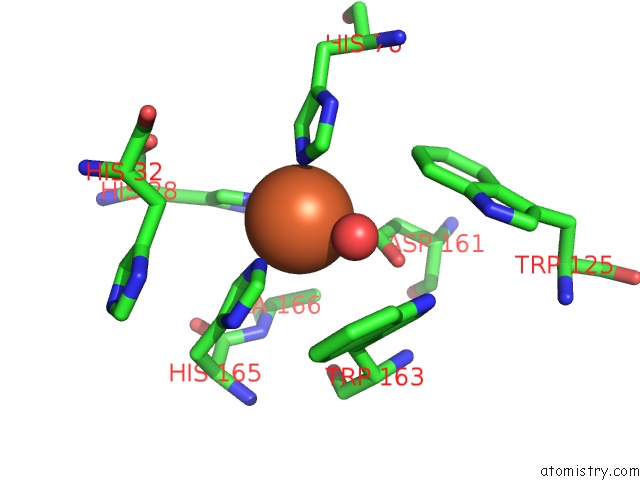
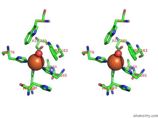
Iron binding site 1 out of 4 in 3esf
Go back to
Iron binding site 1 out
of 4 in the Crystal Structure of the Enzyme Fe-Superoxide Dismutase TBSODB2 From Trypanosoma Brucei
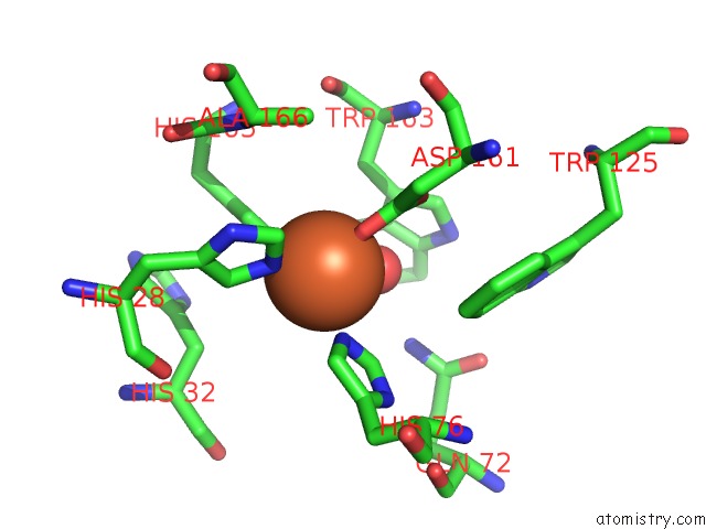
Mono view
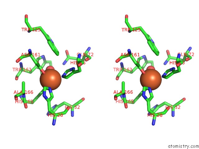
Stereo pair view
Mono view
Stereo pair view
A full contact list of Iron with other atoms in the Fe binding
site number 1 of Crystal Structure of the Enzyme Fe-Superoxide Dismutase TBSODB2 From Trypanosoma Brucei within 5.0Å range:
|
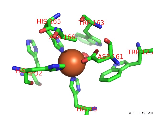
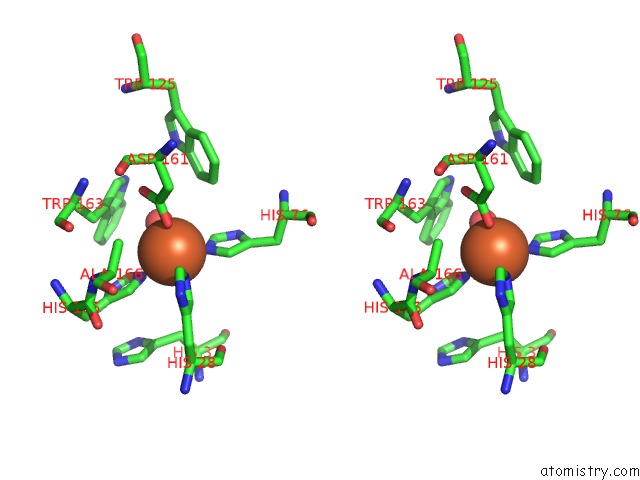
Iron binding site 2 out of 4 in 3esf
Go back to
Iron binding site 2 out
of 4 in the Crystal Structure of the Enzyme Fe-Superoxide Dismutase TBSODB2 From Trypanosoma Brucei
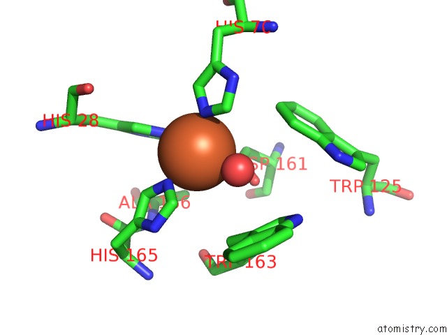
Mono view
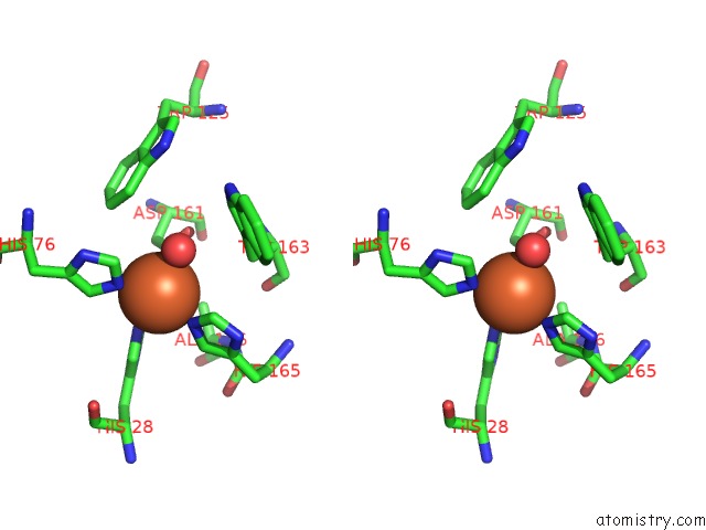
Stereo pair view
Mono view
Stereo pair view
A full contact list of Iron with other atoms in the Fe binding
site number 2 of Crystal Structure of the Enzyme Fe-Superoxide Dismutase TBSODB2 From Trypanosoma Brucei within 5.0Å range:
|
Iron binding site 3 out of 4 in 3esf
Go back to
Iron binding site 3 out
of 4 in the Crystal Structure of the Enzyme Fe-Superoxide Dismutase TBSODB2 From Trypanosoma Brucei
Mono view
Stereo pair view
Mono view
Stereo pair view
A full contact list of Iron with other atoms in the Fe binding
site number 3 of Crystal Structure of the Enzyme Fe-Superoxide Dismutase TBSODB2 From Trypanosoma Brucei within 5.0Å range:
|
Iron binding site 4 out of 4 in 3esf
Go back to
Iron binding site 4 out
of 4 in the Crystal Structure of the Enzyme Fe-Superoxide Dismutase TBSODB2 From Trypanosoma Brucei
Mono view
Stereo pair view
Mono view
Stereo pair view
A full contact list of Iron with other atoms in the Fe binding
site number 4 of Crystal Structure of the Enzyme Fe-Superoxide Dismutase TBSODB2 From Trypanosoma Brucei within 5.0Å range:
|
Reference:
J.F.Bachega,
M.V.Navarro,
L.Bleicher,
R.K.Bortoleto-Bugs,
D.Dive,
P.Hoffmann,
E.Viscogliosi,
R.C.Garratt.
Systematic Structural Studies of Iron Superoxide Dismutases From Human Parasites and A Statistical Coupling Analysis of Metal Binding Specificity Proteins V. 77 26 2009.
ISSN: ISSN 0887-3585
PubMed: 19384994
DOI: 10.1002/PROT.22412
Page generated: Sun Aug 4 09:50:56 2024
ISSN: ISSN 0887-3585
PubMed: 19384994
DOI: 10.1002/PROT.22412
Last articles
Zn in 9J0NZn in 9J0O
Zn in 9J0P
Zn in 9FJX
Zn in 9EKB
Zn in 9C0F
Zn in 9CAH
Zn in 9CH0
Zn in 9CH3
Zn in 9CH1