Iron »
PDB 3etr-3fou »
3fah »
Iron in PDB 3fah: Glycerol Inhibited Form of Aldehyde Oxidoreductase From Desulfovibrio Gigas
Enzymatic activity of Glycerol Inhibited Form of Aldehyde Oxidoreductase From Desulfovibrio Gigas
All present enzymatic activity of Glycerol Inhibited Form of Aldehyde Oxidoreductase From Desulfovibrio Gigas:
1.2.99.7;
1.2.99.7;
Protein crystallography data
The structure of Glycerol Inhibited Form of Aldehyde Oxidoreductase From Desulfovibrio Gigas, PDB code: 3fah
was solved by
T.Santos-Silva,
M.J.Romao,
with X-Ray Crystallography technique. A brief refinement statistics is given in the table below:
| Resolution Low / High (Å) | 26.98 / 1.72 |
| Space group | P 61 2 2 |
| Cell size a, b, c (Å), α, β, γ (°) | 142.560, 142.560, 161.880, 90.00, 90.00, 120.00 |
| R / Rfree (%) | 16 / 19.1 |
Other elements in 3fah:
The structure of Glycerol Inhibited Form of Aldehyde Oxidoreductase From Desulfovibrio Gigas also contains other interesting chemical elements:
| Molybdenum | (Mo) | 1 atom |
| Magnesium | (Mg) | 3 atoms |
| Chlorine | (Cl) | 3 atoms |
Iron Binding Sites:
The binding sites of Iron atom in the Glycerol Inhibited Form of Aldehyde Oxidoreductase From Desulfovibrio Gigas
(pdb code 3fah). This binding sites where shown within
5.0 Angstroms radius around Iron atom.
In total 4 binding sites of Iron where determined in the Glycerol Inhibited Form of Aldehyde Oxidoreductase From Desulfovibrio Gigas, PDB code: 3fah:
Jump to Iron binding site number: 1; 2; 3; 4;
In total 4 binding sites of Iron where determined in the Glycerol Inhibited Form of Aldehyde Oxidoreductase From Desulfovibrio Gigas, PDB code: 3fah:
Jump to Iron binding site number: 1; 2; 3; 4;
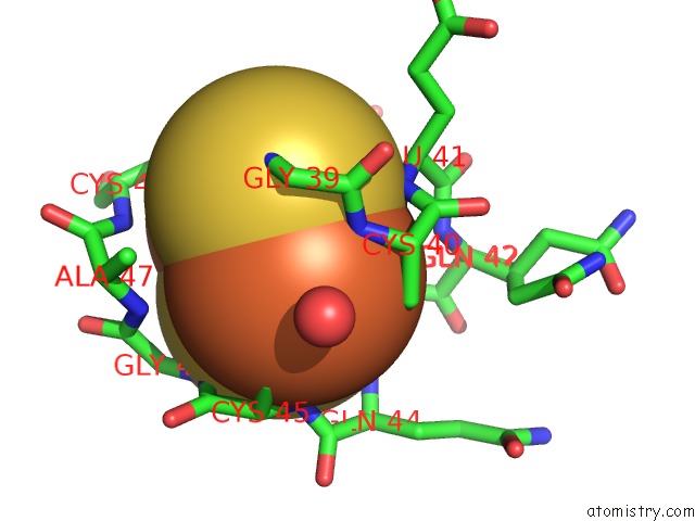
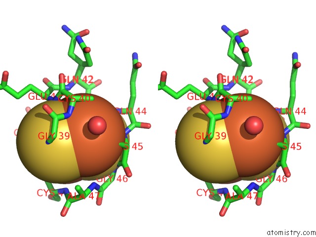
Iron binding site 1 out of 4 in 3fah
Go back to
Iron binding site 1 out
of 4 in the Glycerol Inhibited Form of Aldehyde Oxidoreductase From Desulfovibrio Gigas
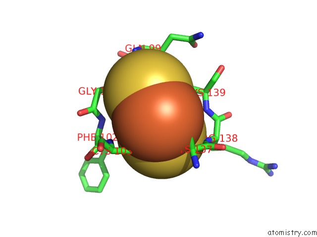
Mono view
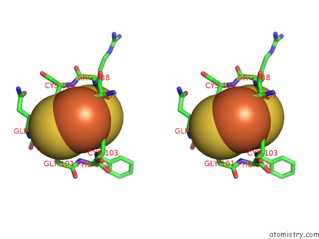
Stereo pair view
Mono view
Stereo pair view
A full contact list of Iron with other atoms in the Fe binding
site number 1 of Glycerol Inhibited Form of Aldehyde Oxidoreductase From Desulfovibrio Gigas within 5.0Å range:
|
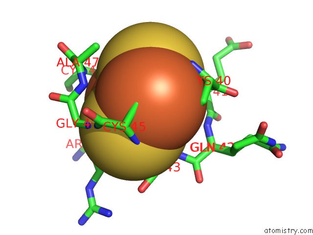
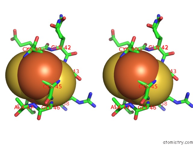
Iron binding site 2 out of 4 in 3fah
Go back to
Iron binding site 2 out
of 4 in the Glycerol Inhibited Form of Aldehyde Oxidoreductase From Desulfovibrio Gigas
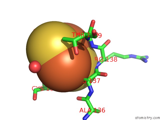
Mono view
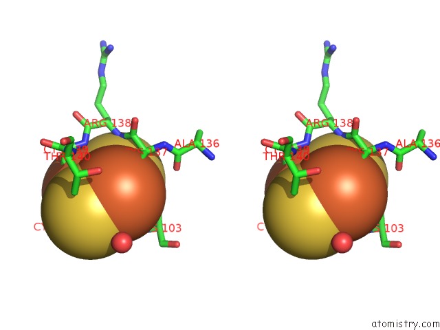
Stereo pair view
Mono view
Stereo pair view
A full contact list of Iron with other atoms in the Fe binding
site number 2 of Glycerol Inhibited Form of Aldehyde Oxidoreductase From Desulfovibrio Gigas within 5.0Å range:
|
Iron binding site 3 out of 4 in 3fah
Go back to
Iron binding site 3 out
of 4 in the Glycerol Inhibited Form of Aldehyde Oxidoreductase From Desulfovibrio Gigas
Mono view
Stereo pair view
Mono view
Stereo pair view
A full contact list of Iron with other atoms in the Fe binding
site number 3 of Glycerol Inhibited Form of Aldehyde Oxidoreductase From Desulfovibrio Gigas within 5.0Å range:
|
Iron binding site 4 out of 4 in 3fah
Go back to
Iron binding site 4 out
of 4 in the Glycerol Inhibited Form of Aldehyde Oxidoreductase From Desulfovibrio Gigas
Mono view
Stereo pair view
Mono view
Stereo pair view
A full contact list of Iron with other atoms in the Fe binding
site number 4 of Glycerol Inhibited Form of Aldehyde Oxidoreductase From Desulfovibrio Gigas within 5.0Å range:
|
Reference:
T.Santos-Silva,
F.Ferroni,
A.Thapper,
J.Marangon,
P.J.Gonzalez,
A.C.Rizzi,
I.Moura,
J.J.Moura,
M.J.Romao,
C.D.Brondino.
Kinetic, Structural, and Epr Studies Reveal That Aldehyde Oxidoreductase From Desulfovibrio Gigas Does Not Need A Sulfido Ligand For Catalysis and Give Evidence For A Direct Mo-C Interaction in A Biological System. J.Am.Chem.Soc. V. 131 7990 2009.
ISSN: ISSN 0002-7863
PubMed: 19459677
DOI: 10.1021/JA809448R
Page generated: Sun Aug 4 09:58:06 2024
ISSN: ISSN 0002-7863
PubMed: 19459677
DOI: 10.1021/JA809448R
Last articles
Zn in 9MJ5Zn in 9HNW
Zn in 9G0L
Zn in 9FNE
Zn in 9DZN
Zn in 9E0I
Zn in 9D32
Zn in 9DAK
Zn in 8ZXC
Zn in 8ZUF