Iron »
PDB 3etr-3fou »
3fmq »
Iron in PDB 3fmq: Crystal Structure of An Encephalitozoon Cuniculi Methionine Aminopeptidase Type 2 with Angiogenesis Inhibitor Fumagillin Bound
Enzymatic activity of Crystal Structure of An Encephalitozoon Cuniculi Methionine Aminopeptidase Type 2 with Angiogenesis Inhibitor Fumagillin Bound
All present enzymatic activity of Crystal Structure of An Encephalitozoon Cuniculi Methionine Aminopeptidase Type 2 with Angiogenesis Inhibitor Fumagillin Bound:
3.4.11.18;
3.4.11.18;
Protein crystallography data
The structure of Crystal Structure of An Encephalitozoon Cuniculi Methionine Aminopeptidase Type 2 with Angiogenesis Inhibitor Fumagillin Bound, PDB code: 3fmq
was solved by
J.J.Alvarado,
M.Russell,
A.Zhang,
J.Adams,
R.Toro,
S.K.Burley,
L.M.Weiss,
S.C.Almo,
New York Sgx Research Center For Structural Genomics(Nysgxrc),
with X-Ray Crystallography technique. A brief refinement statistics is given in the table below:
| Resolution Low / High (Å) | 50.00 / 2.50 |
| Space group | P 1 21 1 |
| Cell size a, b, c (Å), α, β, γ (°) | 62.008, 94.238, 66.325, 90.00, 99.43, 90.00 |
| R / Rfree (%) | 17.3 / 22.9 |
Iron Binding Sites:
The binding sites of Iron atom in the Crystal Structure of An Encephalitozoon Cuniculi Methionine Aminopeptidase Type 2 with Angiogenesis Inhibitor Fumagillin Bound
(pdb code 3fmq). This binding sites where shown within
5.0 Angstroms radius around Iron atom.
In total 4 binding sites of Iron where determined in the Crystal Structure of An Encephalitozoon Cuniculi Methionine Aminopeptidase Type 2 with Angiogenesis Inhibitor Fumagillin Bound, PDB code: 3fmq:
Jump to Iron binding site number: 1; 2; 3; 4;
In total 4 binding sites of Iron where determined in the Crystal Structure of An Encephalitozoon Cuniculi Methionine Aminopeptidase Type 2 with Angiogenesis Inhibitor Fumagillin Bound, PDB code: 3fmq:
Jump to Iron binding site number: 1; 2; 3; 4;
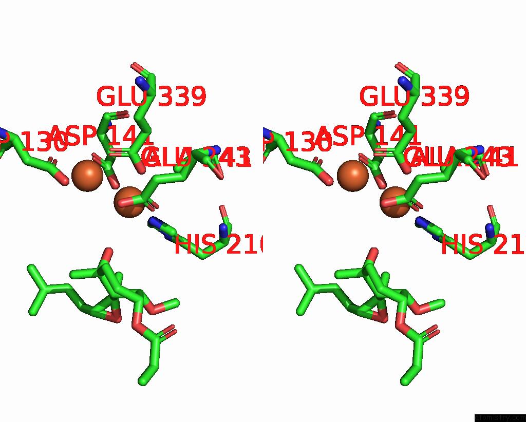
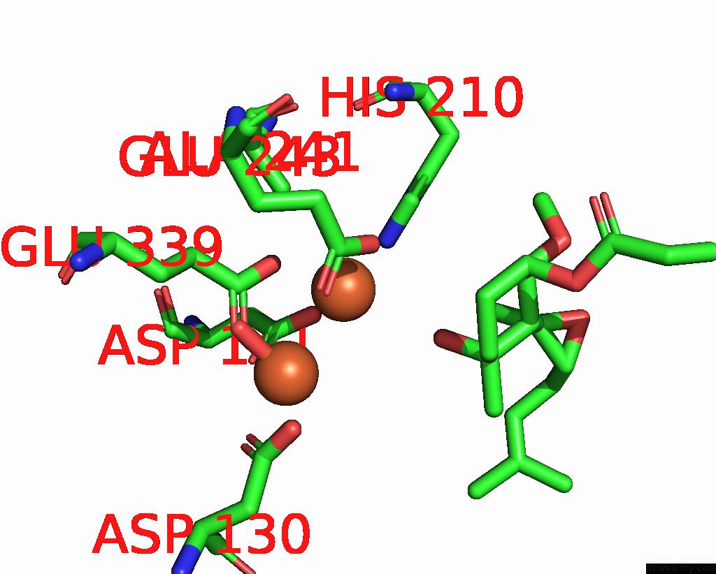
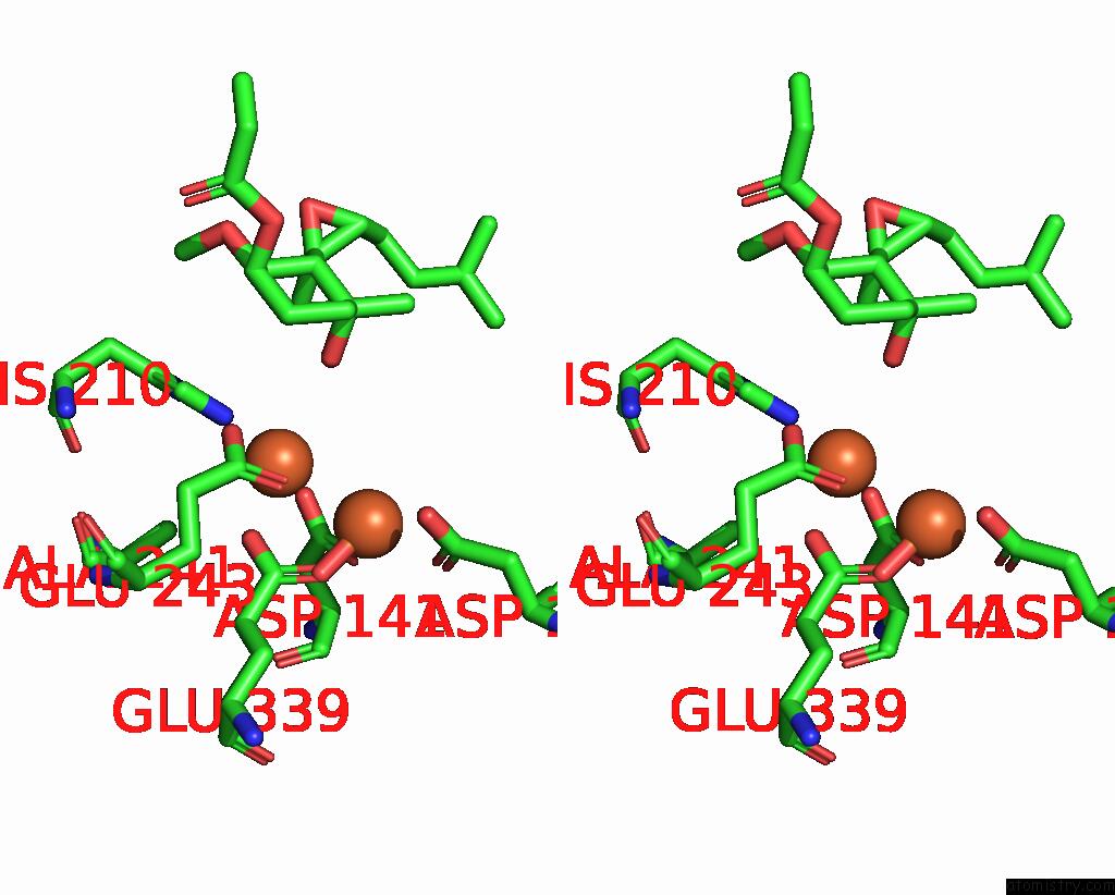
Iron binding site 1 out of 4 in 3fmq
Go back to
Iron binding site 1 out
of 4 in the Crystal Structure of An Encephalitozoon Cuniculi Methionine Aminopeptidase Type 2 with Angiogenesis Inhibitor Fumagillin Bound
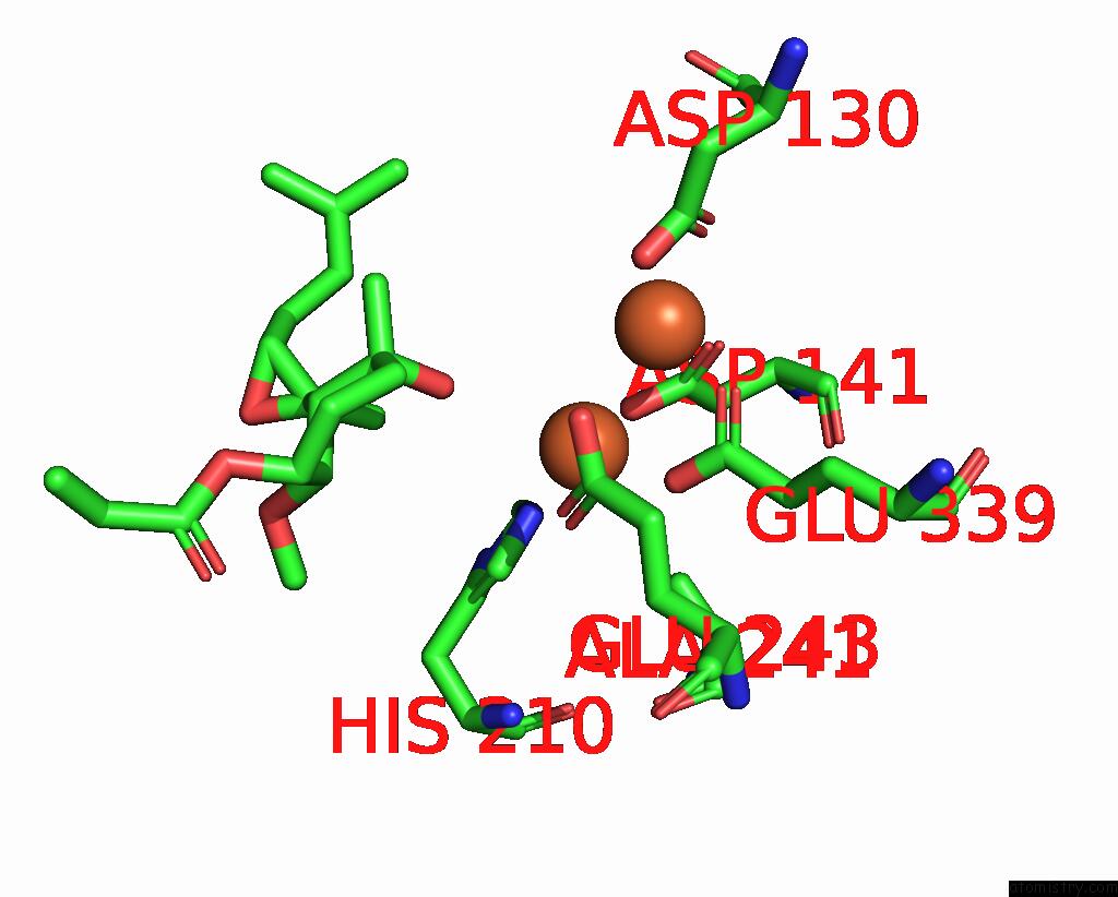
Mono view
Stereo pair view
Mono view
Stereo pair view
A full contact list of Iron with other atoms in the Fe binding
site number 1 of Crystal Structure of An Encephalitozoon Cuniculi Methionine Aminopeptidase Type 2 with Angiogenesis Inhibitor Fumagillin Bound within 5.0Å range:
|
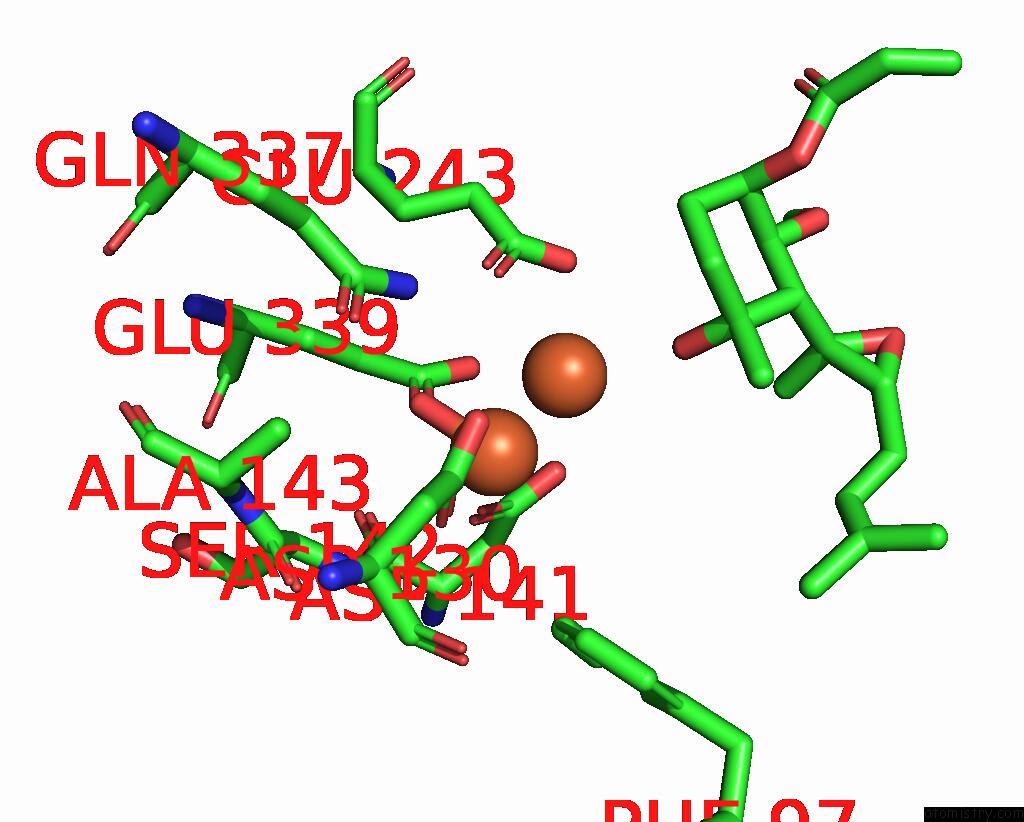
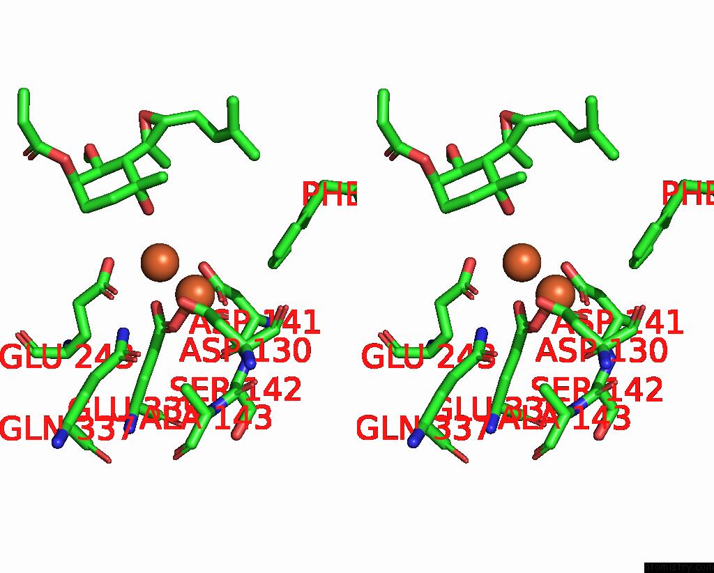
Iron binding site 2 out of 4 in 3fmq
Go back to
Iron binding site 2 out
of 4 in the Crystal Structure of An Encephalitozoon Cuniculi Methionine Aminopeptidase Type 2 with Angiogenesis Inhibitor Fumagillin Bound
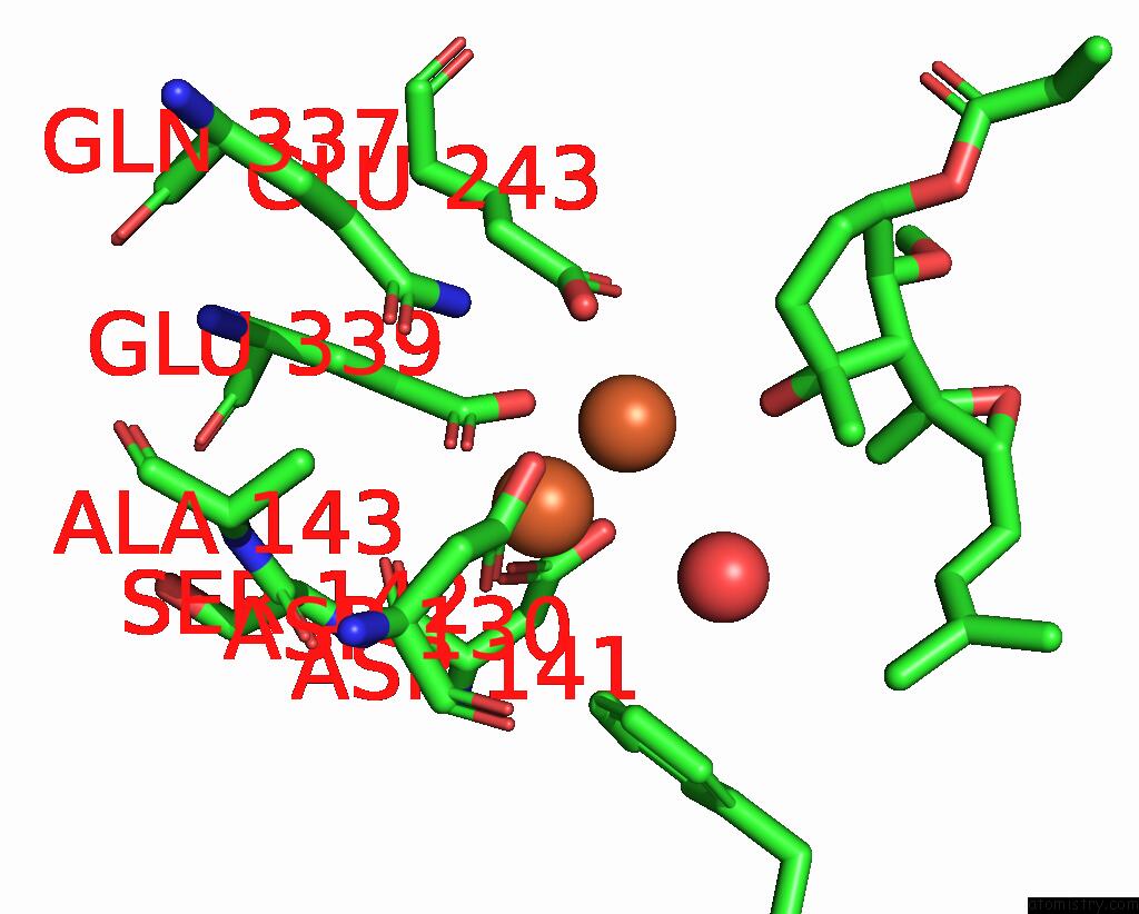
Mono view
Stereo pair view
Mono view
Stereo pair view
A full contact list of Iron with other atoms in the Fe binding
site number 2 of Crystal Structure of An Encephalitozoon Cuniculi Methionine Aminopeptidase Type 2 with Angiogenesis Inhibitor Fumagillin Bound within 5.0Å range:
|
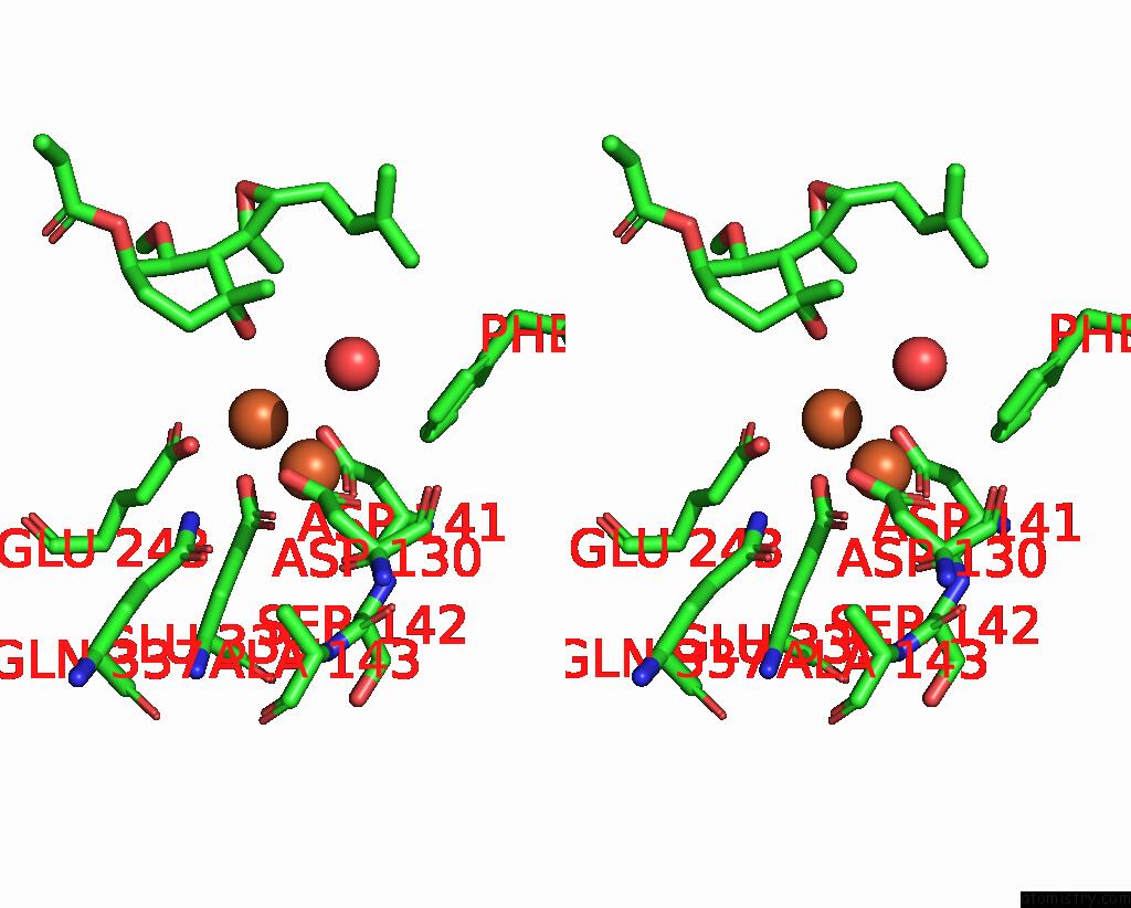
Iron binding site 3 out of 4 in 3fmq
Go back to
Iron binding site 3 out
of 4 in the Crystal Structure of An Encephalitozoon Cuniculi Methionine Aminopeptidase Type 2 with Angiogenesis Inhibitor Fumagillin Bound
Mono view
Stereo pair view
Mono view
Stereo pair view
A full contact list of Iron with other atoms in the Fe binding
site number 3 of Crystal Structure of An Encephalitozoon Cuniculi Methionine Aminopeptidase Type 2 with Angiogenesis Inhibitor Fumagillin Bound within 5.0Å range:
|
Iron binding site 4 out of 4 in 3fmq
Go back to
Iron binding site 4 out
of 4 in the Crystal Structure of An Encephalitozoon Cuniculi Methionine Aminopeptidase Type 2 with Angiogenesis Inhibitor Fumagillin Bound
Mono view
Stereo pair view
Mono view
Stereo pair view
A full contact list of Iron with other atoms in the Fe binding
site number 4 of Crystal Structure of An Encephalitozoon Cuniculi Methionine Aminopeptidase Type 2 with Angiogenesis Inhibitor Fumagillin Bound within 5.0Å range:
|
Reference:
J.J.Alvarado,
A.Nemkal,
J.M.Sauder,
M.Russell,
D.E.Akiyoshi,
W.Shi,
S.C.Almo,
L.M.Weiss.
Structure of A Microsporidian Methionine Aminopeptidase Type 2 Complexed with Fumagillin and Tnp-470. Mol.Biochem.Parasitol. V. 168 158 2009.
ISSN: ISSN 0166-6851
PubMed: 19660503
DOI: 10.1016/J.MOLBIOPARA.2009.07.008
Page generated: Sun Aug 4 10:07:31 2024
ISSN: ISSN 0166-6851
PubMed: 19660503
DOI: 10.1016/J.MOLBIOPARA.2009.07.008
Last articles
Zn in 9MJ5Zn in 9HNW
Zn in 9G0L
Zn in 9FNE
Zn in 9DZN
Zn in 9E0I
Zn in 9D32
Zn in 9DAK
Zn in 8ZXC
Zn in 8ZUF