Iron »
PDB 3fpv-3gcj »
3fwy »
Iron in PDB 3fwy: Crystal Structure of the L Protein of Rhodobacter Sphaeroides Light- Independent Protochlorophyllide Reductase (Bchl) with Mgadp Bound: A Homologue of the Nitrogenase Fe Protein
Protein crystallography data
The structure of Crystal Structure of the L Protein of Rhodobacter Sphaeroides Light- Independent Protochlorophyllide Reductase (Bchl) with Mgadp Bound: A Homologue of the Nitrogenase Fe Protein, PDB code: 3fwy
was solved by
R.Sarma,
B.M.Barney,
T.L.Hamilton,
A.Jones,
L.C.Seefeldt,
J.W.Peters,
with X-Ray Crystallography technique. A brief refinement statistics is given in the table below:
| Resolution Low / High (Å) | 33.02 / 1.63 |
| Space group | P 21 21 21 |
| Cell size a, b, c (Å), α, β, γ (°) | 56.729, 86.622, 117.169, 90.00, 90.00, 90.00 |
| R / Rfree (%) | 17.4 / 19.9 |
Other elements in 3fwy:
The structure of Crystal Structure of the L Protein of Rhodobacter Sphaeroides Light- Independent Protochlorophyllide Reductase (Bchl) with Mgadp Bound: A Homologue of the Nitrogenase Fe Protein also contains other interesting chemical elements:
| Magnesium | (Mg) | 2 atoms |
Iron Binding Sites:
The binding sites of Iron atom in the Crystal Structure of the L Protein of Rhodobacter Sphaeroides Light- Independent Protochlorophyllide Reductase (Bchl) with Mgadp Bound: A Homologue of the Nitrogenase Fe Protein
(pdb code 3fwy). This binding sites where shown within
5.0 Angstroms radius around Iron atom.
In total 4 binding sites of Iron where determined in the Crystal Structure of the L Protein of Rhodobacter Sphaeroides Light- Independent Protochlorophyllide Reductase (Bchl) with Mgadp Bound: A Homologue of the Nitrogenase Fe Protein, PDB code: 3fwy:
Jump to Iron binding site number: 1; 2; 3; 4;
In total 4 binding sites of Iron where determined in the Crystal Structure of the L Protein of Rhodobacter Sphaeroides Light- Independent Protochlorophyllide Reductase (Bchl) with Mgadp Bound: A Homologue of the Nitrogenase Fe Protein, PDB code: 3fwy:
Jump to Iron binding site number: 1; 2; 3; 4;
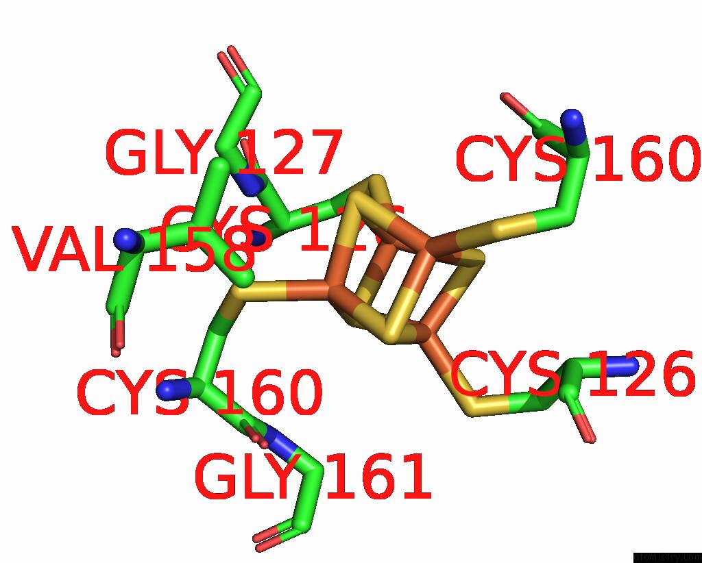
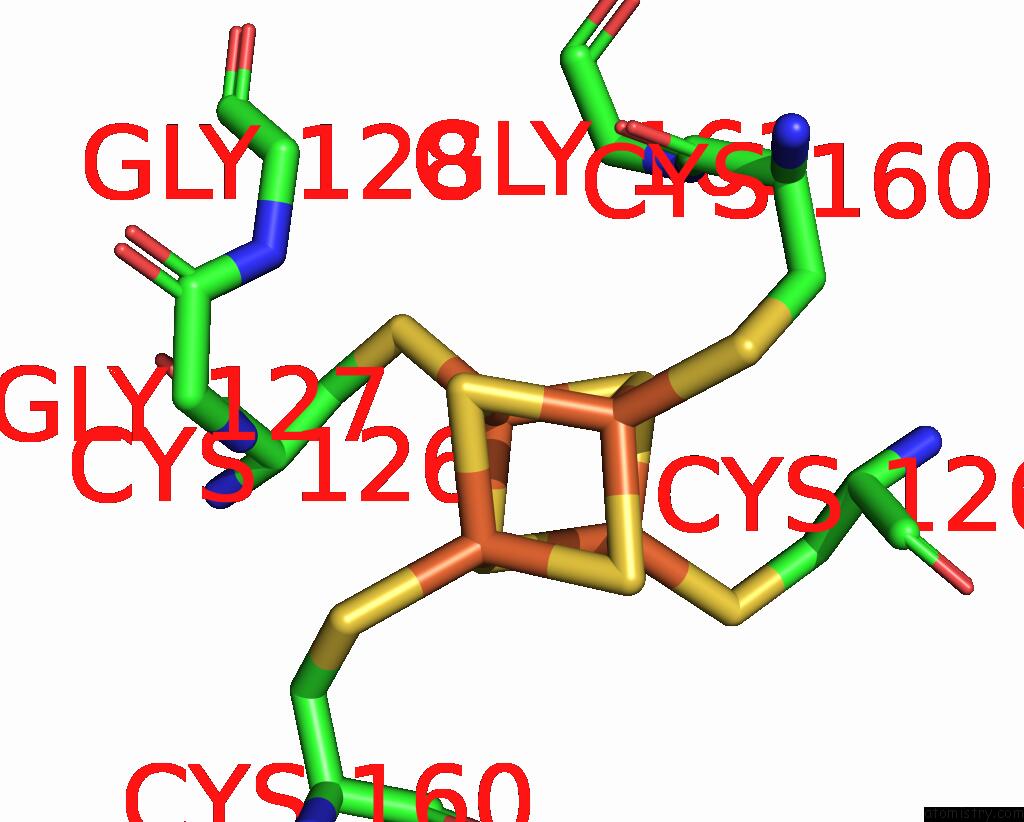
Iron binding site 1 out of 4 in 3fwy
Go back to
Iron binding site 1 out
of 4 in the Crystal Structure of the L Protein of Rhodobacter Sphaeroides Light- Independent Protochlorophyllide Reductase (Bchl) with Mgadp Bound: A Homologue of the Nitrogenase Fe Protein
Mono view
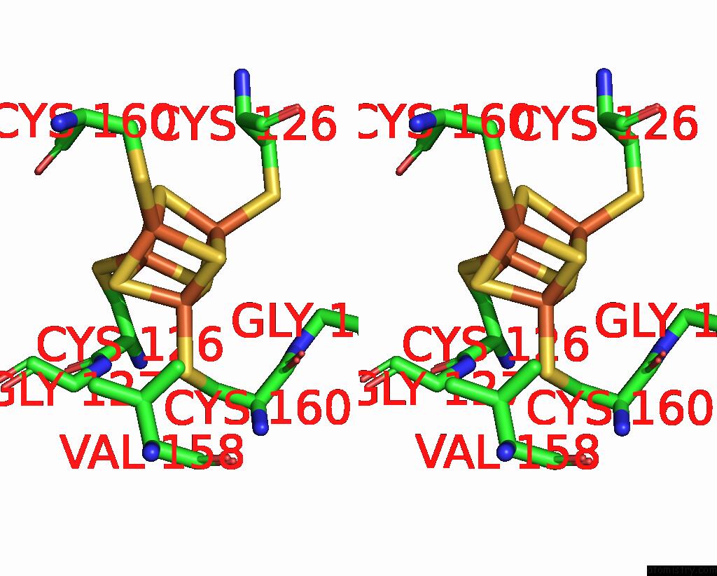
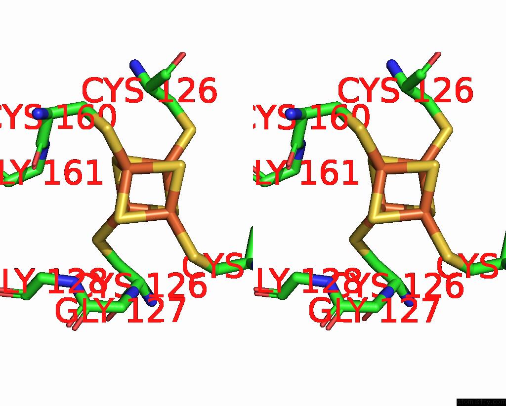
Stereo pair view
Mono view
Stereo pair view
A full contact list of Iron with other atoms in the Fe binding
site number 1 of Crystal Structure of the L Protein of Rhodobacter Sphaeroides Light- Independent Protochlorophyllide Reductase (Bchl) with Mgadp Bound: A Homologue of the Nitrogenase Fe Protein within 5.0Å range:
|
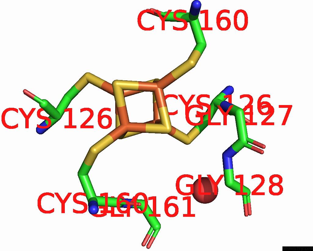
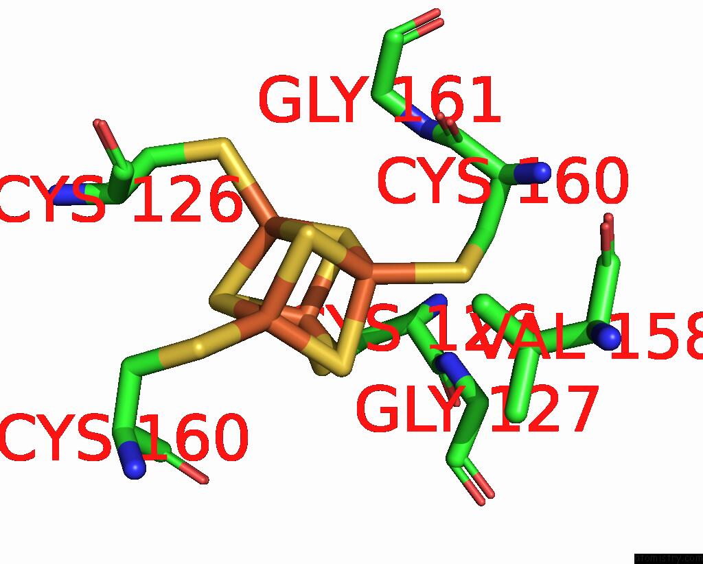
Iron binding site 2 out of 4 in 3fwy
Go back to
Iron binding site 2 out
of 4 in the Crystal Structure of the L Protein of Rhodobacter Sphaeroides Light- Independent Protochlorophyllide Reductase (Bchl) with Mgadp Bound: A Homologue of the Nitrogenase Fe Protein
Mono view
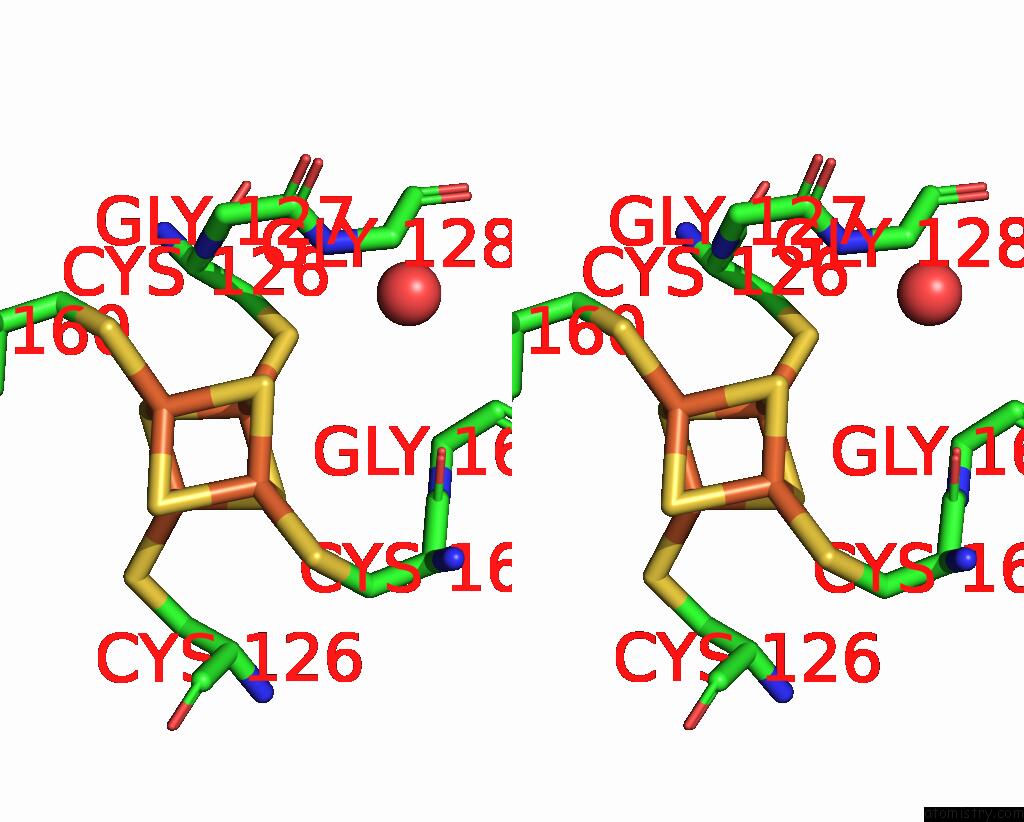
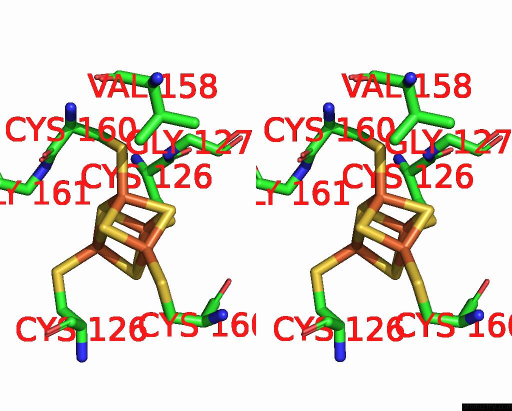
Stereo pair view
Mono view
Stereo pair view
A full contact list of Iron with other atoms in the Fe binding
site number 2 of Crystal Structure of the L Protein of Rhodobacter Sphaeroides Light- Independent Protochlorophyllide Reductase (Bchl) with Mgadp Bound: A Homologue of the Nitrogenase Fe Protein within 5.0Å range:
|
Iron binding site 3 out of 4 in 3fwy
Go back to
Iron binding site 3 out
of 4 in the Crystal Structure of the L Protein of Rhodobacter Sphaeroides Light- Independent Protochlorophyllide Reductase (Bchl) with Mgadp Bound: A Homologue of the Nitrogenase Fe Protein
Mono view
Stereo pair view
Mono view
Stereo pair view
A full contact list of Iron with other atoms in the Fe binding
site number 3 of Crystal Structure of the L Protein of Rhodobacter Sphaeroides Light- Independent Protochlorophyllide Reductase (Bchl) with Mgadp Bound: A Homologue of the Nitrogenase Fe Protein within 5.0Å range:
|
Iron binding site 4 out of 4 in 3fwy
Go back to
Iron binding site 4 out
of 4 in the Crystal Structure of the L Protein of Rhodobacter Sphaeroides Light- Independent Protochlorophyllide Reductase (Bchl) with Mgadp Bound: A Homologue of the Nitrogenase Fe Protein
Mono view
Stereo pair view
Mono view
Stereo pair view
A full contact list of Iron with other atoms in the Fe binding
site number 4 of Crystal Structure of the L Protein of Rhodobacter Sphaeroides Light- Independent Protochlorophyllide Reductase (Bchl) with Mgadp Bound: A Homologue of the Nitrogenase Fe Protein within 5.0Å range:
|
Reference:
R.Sarma,
B.M.Barney,
T.L.Hamilton,
A.Jones,
L.C.Seefeldt,
J.W.Peters.
Crystal Structure of the L Protein of Rhodobacter Sphaeroides Light-Independent Protochlorophyllide Reductase with Mgadp Bound: A Homologue of the Nitrogenase Fe Protein. Biochemistry V. 47 13004 2008.
ISSN: ISSN 0006-2960
PubMed: 19006326
DOI: 10.1021/BI801058R
Page generated: Sun Aug 4 10:20:39 2024
ISSN: ISSN 0006-2960
PubMed: 19006326
DOI: 10.1021/BI801058R
Last articles
F in 7QMYF in 7QMX
F in 7QMW
F in 7QMZ
F in 7QMU
F in 7QMV
F in 7QMT
F in 7QMS
F in 7QMQ
F in 7QMR