Iron »
PDB 3gck-3h33 »
3git »
Iron in PDB 3git: Crystal Structure of A Truncated Acetyl-Coa Synthase
Enzymatic activity of Crystal Structure of A Truncated Acetyl-Coa Synthase
All present enzymatic activity of Crystal Structure of A Truncated Acetyl-Coa Synthase:
2.3.1.169;
2.3.1.169;
Protein crystallography data
The structure of Crystal Structure of A Truncated Acetyl-Coa Synthase, PDB code: 3git
was solved by
A.Volbeda,
C.Darnault,
J.C.Fontecilla-Camps,
with X-Ray Crystallography technique. A brief refinement statistics is given in the table below:
| Resolution Low / High (Å) | 19.98 / 3.00 |
| Space group | P 31 2 1 |
| Cell size a, b, c (Å), α, β, γ (°) | 166.400, 166.400, 245.200, 90.00, 90.00, 120.00 |
| R / Rfree (%) | 17.1 / 20.8 |
Other elements in 3git:
The structure of Crystal Structure of A Truncated Acetyl-Coa Synthase also contains other interesting chemical elements:
| Zinc | (Zn) | 6 atoms |
Iron Binding Sites:
Pages:
>>> Page 1 <<< Page 2, Binding sites: 11 - 20; Page 3, Binding sites: 21 - 24;Binding sites:
The binding sites of Iron atom in the Crystal Structure of A Truncated Acetyl-Coa Synthase (pdb code 3git). This binding sites where shown within 5.0 Angstroms radius around Iron atom.In total 24 binding sites of Iron where determined in the Crystal Structure of A Truncated Acetyl-Coa Synthase, PDB code: 3git:
Jump to Iron binding site number: 1; 2; 3; 4; 5; 6; 7; 8; 9; 10;
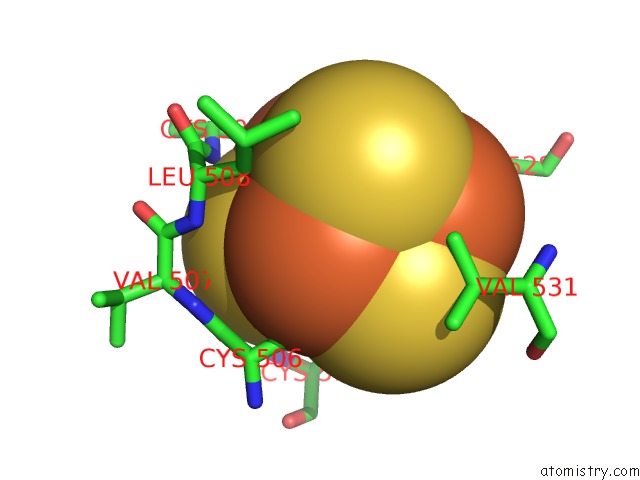
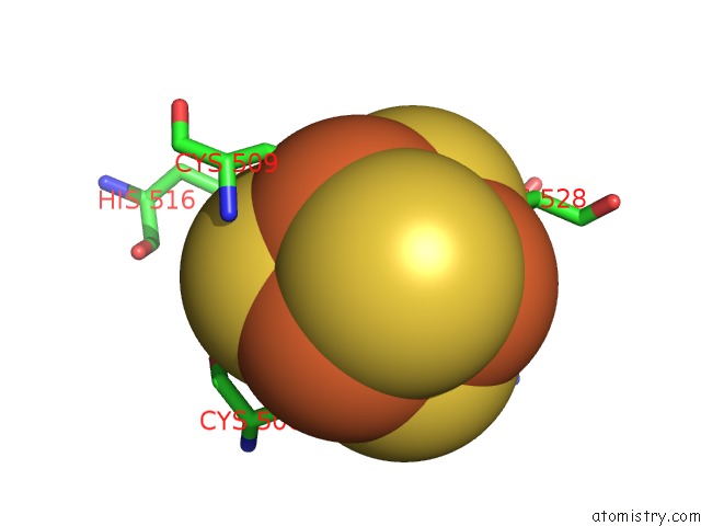
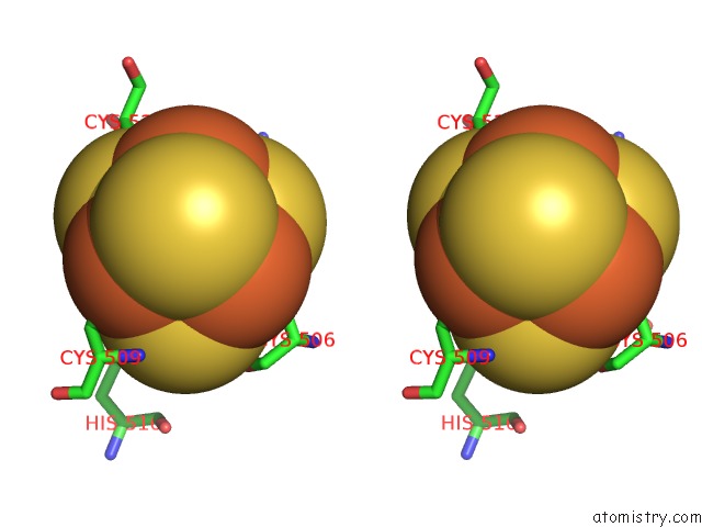
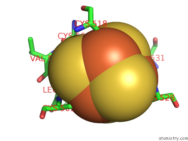
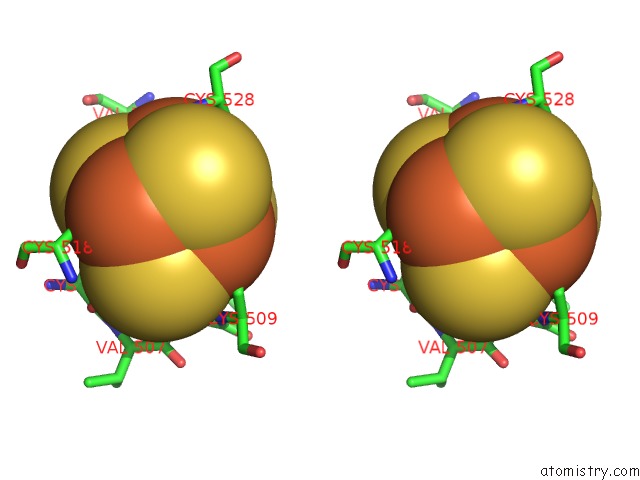
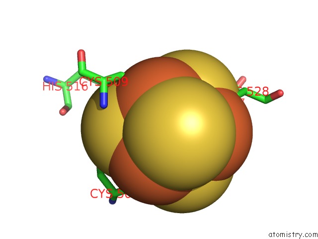
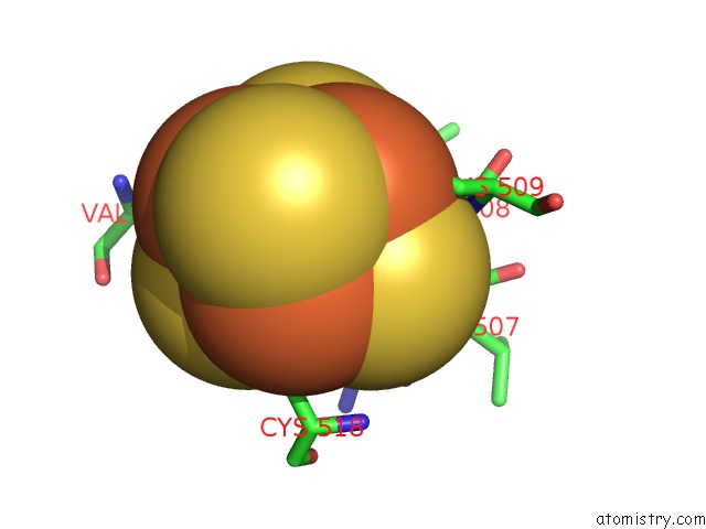
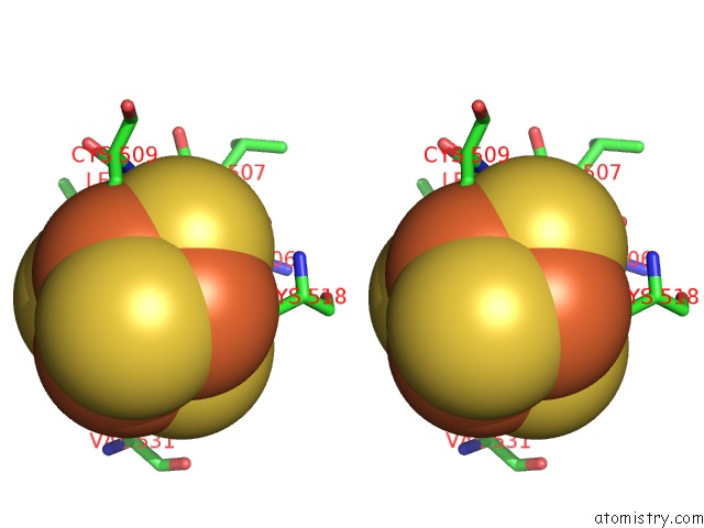
Iron binding site 1 out of 24 in 3git
Go back to
Iron binding site 1 out
of 24 in the Crystal Structure of A Truncated Acetyl-Coa Synthase
Mono view
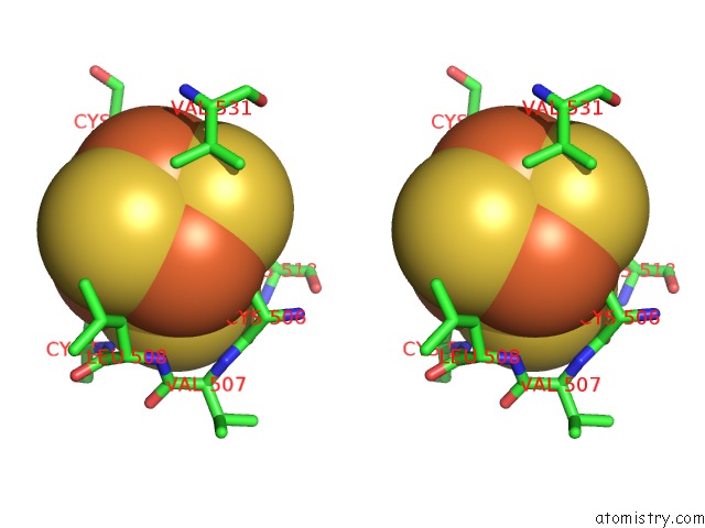
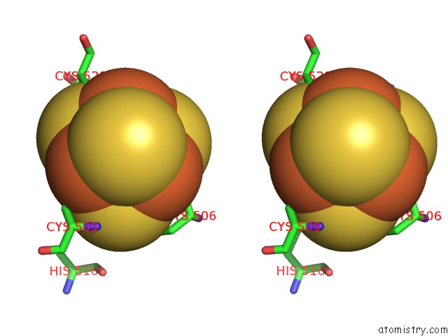
Stereo pair view
Mono view
Stereo pair view
A full contact list of Iron with other atoms in the Fe binding
site number 1 of Crystal Structure of A Truncated Acetyl-Coa Synthase within 5.0Å range:
|
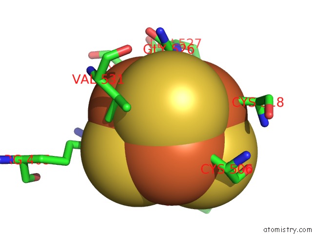
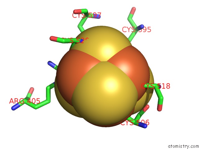
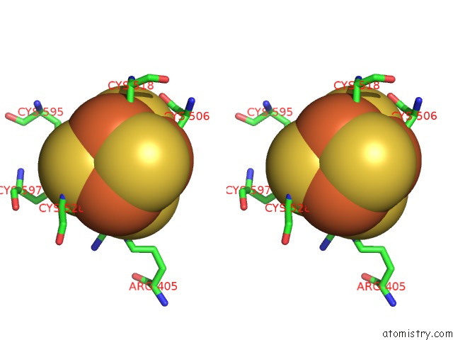
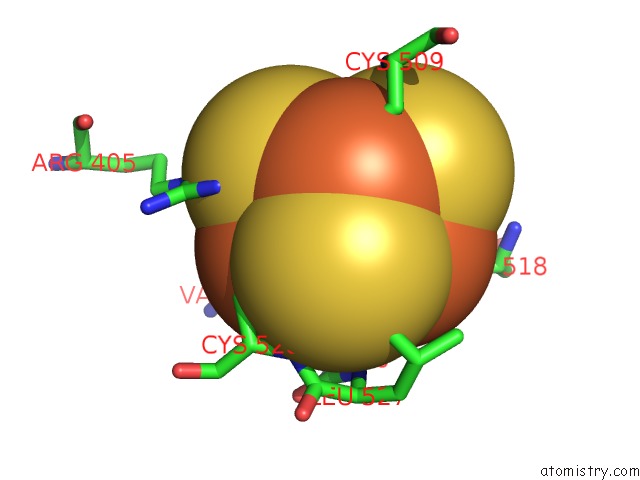
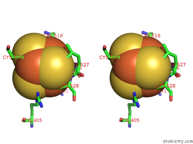
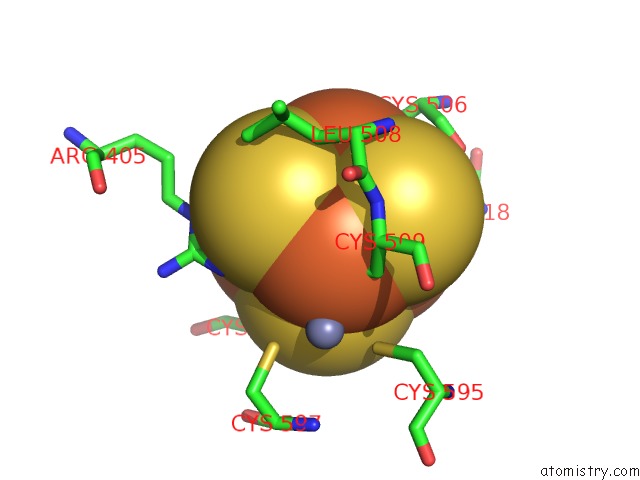
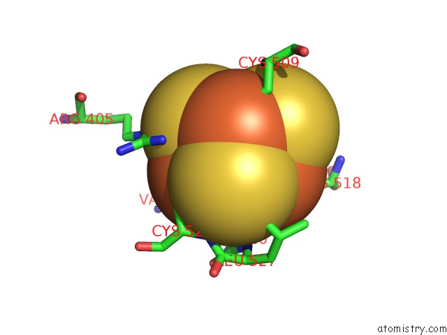
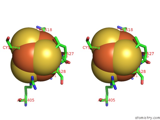
Iron binding site 2 out of 24 in 3git
Go back to
Iron binding site 2 out
of 24 in the Crystal Structure of A Truncated Acetyl-Coa Synthase
Mono view
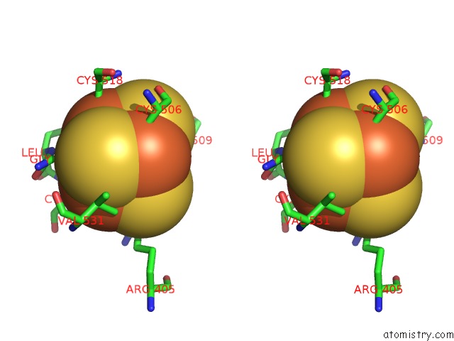
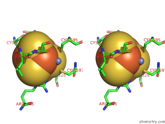
Stereo pair view
Mono view
Stereo pair view
A full contact list of Iron with other atoms in the Fe binding
site number 2 of Crystal Structure of A Truncated Acetyl-Coa Synthase within 5.0Å range:
|
Iron binding site 3 out of 24 in 3git
Go back to
Iron binding site 3 out
of 24 in the Crystal Structure of A Truncated Acetyl-Coa Synthase
Mono view
Stereo pair view
Mono view
Stereo pair view
A full contact list of Iron with other atoms in the Fe binding
site number 3 of Crystal Structure of A Truncated Acetyl-Coa Synthase within 5.0Å range:
|
Iron binding site 4 out of 24 in 3git
Go back to
Iron binding site 4 out
of 24 in the Crystal Structure of A Truncated Acetyl-Coa Synthase
Mono view
Stereo pair view
Mono view
Stereo pair view
A full contact list of Iron with other atoms in the Fe binding
site number 4 of Crystal Structure of A Truncated Acetyl-Coa Synthase within 5.0Å range:
|
Iron binding site 5 out of 24 in 3git
Go back to
Iron binding site 5 out
of 24 in the Crystal Structure of A Truncated Acetyl-Coa Synthase
Mono view
Stereo pair view
Mono view
Stereo pair view
A full contact list of Iron with other atoms in the Fe binding
site number 5 of Crystal Structure of A Truncated Acetyl-Coa Synthase within 5.0Å range:
|
Iron binding site 6 out of 24 in 3git
Go back to
Iron binding site 6 out
of 24 in the Crystal Structure of A Truncated Acetyl-Coa Synthase
Mono view
Stereo pair view
Mono view
Stereo pair view
A full contact list of Iron with other atoms in the Fe binding
site number 6 of Crystal Structure of A Truncated Acetyl-Coa Synthase within 5.0Å range:
|
Iron binding site 7 out of 24 in 3git
Go back to
Iron binding site 7 out
of 24 in the Crystal Structure of A Truncated Acetyl-Coa Synthase
Mono view
Stereo pair view
Mono view
Stereo pair view
A full contact list of Iron with other atoms in the Fe binding
site number 7 of Crystal Structure of A Truncated Acetyl-Coa Synthase within 5.0Å range:
|
Iron binding site 8 out of 24 in 3git
Go back to
Iron binding site 8 out
of 24 in the Crystal Structure of A Truncated Acetyl-Coa Synthase
Mono view
Stereo pair view
Mono view
Stereo pair view
A full contact list of Iron with other atoms in the Fe binding
site number 8 of Crystal Structure of A Truncated Acetyl-Coa Synthase within 5.0Å range:
|
Iron binding site 9 out of 24 in 3git
Go back to
Iron binding site 9 out
of 24 in the Crystal Structure of A Truncated Acetyl-Coa Synthase
Mono view
Stereo pair view
Mono view
Stereo pair view
A full contact list of Iron with other atoms in the Fe binding
site number 9 of Crystal Structure of A Truncated Acetyl-Coa Synthase within 5.0Å range:
|
Iron binding site 10 out of 24 in 3git
Go back to
Iron binding site 10 out
of 24 in the Crystal Structure of A Truncated Acetyl-Coa Synthase
Mono view
Stereo pair view
Mono view
Stereo pair view
A full contact list of Iron with other atoms in the Fe binding
site number 10 of Crystal Structure of A Truncated Acetyl-Coa Synthase within 5.0Å range:
|
Reference:
A.Volbeda,
C.Darnault,
X.Tan,
P.A.Lindahl,
J.C.Fontecilla-Camps.
Novel Domain Arrangement in the Crystal Structure of A Truncated Acetyl-Coa Synthase From Moorella Thermoacetica Biochemistry V. 48 7916 2009.
ISSN: ISSN 0006-2960
PubMed: 19650626
DOI: 10.1021/BI9003952
Page generated: Sun Aug 4 10:42:00 2024
ISSN: ISSN 0006-2960
PubMed: 19650626
DOI: 10.1021/BI9003952
Last articles
F in 4IF4F in 4IIZ
F in 4IDQ
F in 4IGH
F in 4IGA
F in 4IBJ
F in 4IFY
F in 4IFV
F in 4IDO
F in 4ICC