Iron »
PDB 3gck-3h33 »
3gqg »
Iron in PDB 3gqg: Crystal Structure at Acidic pH of the Ferric Form of the Root Effect Hemoglobin From Trematomus Bernacchii.
Protein crystallography data
The structure of Crystal Structure at Acidic pH of the Ferric Form of the Root Effect Hemoglobin From Trematomus Bernacchii., PDB code: 3gqg
was solved by
A.Vergara,
M.Franzese,
A.Merlino,
G.Bonomi,
L.Mazzarella,
with X-Ray Crystallography technique. A brief refinement statistics is given in the table below:
| Resolution Low / High (Å) | 13.90 / 1.73 |
| Space group | P 1 21 1 |
| Cell size a, b, c (Å), α, β, γ (°) | 61.720, 94.780, 61.720, 90.00, 90.09, 90.00 |
| R / Rfree (%) | 15.6 / 19.7 |
Iron Binding Sites:
The binding sites of Iron atom in the Crystal Structure at Acidic pH of the Ferric Form of the Root Effect Hemoglobin From Trematomus Bernacchii.
(pdb code 3gqg). This binding sites where shown within
5.0 Angstroms radius around Iron atom.
In total 4 binding sites of Iron where determined in the Crystal Structure at Acidic pH of the Ferric Form of the Root Effect Hemoglobin From Trematomus Bernacchii., PDB code: 3gqg:
Jump to Iron binding site number: 1; 2; 3; 4;
In total 4 binding sites of Iron where determined in the Crystal Structure at Acidic pH of the Ferric Form of the Root Effect Hemoglobin From Trematomus Bernacchii., PDB code: 3gqg:
Jump to Iron binding site number: 1; 2; 3; 4;
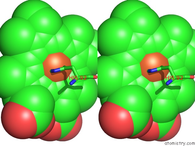
Iron binding site 1 out of 4 in 3gqg
Go back to
Iron binding site 1 out
of 4 in the Crystal Structure at Acidic pH of the Ferric Form of the Root Effect Hemoglobin From Trematomus Bernacchii.
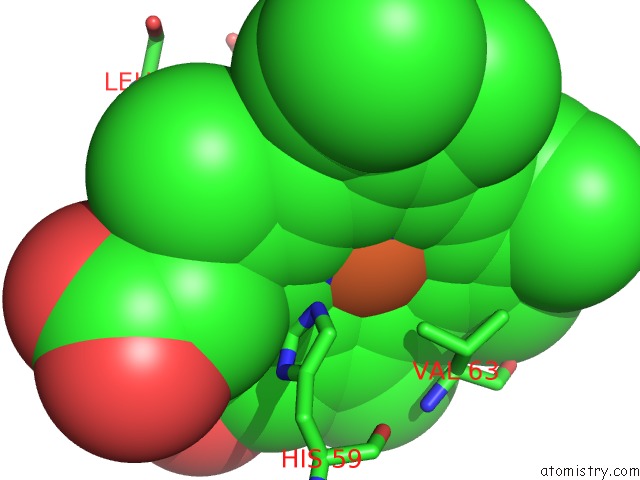
Mono view
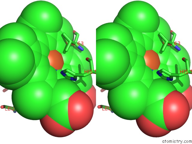
Stereo pair view
Mono view
Stereo pair view
A full contact list of Iron with other atoms in the Fe binding
site number 1 of Crystal Structure at Acidic pH of the Ferric Form of the Root Effect Hemoglobin From Trematomus Bernacchii. within 5.0Å range:
|
Iron binding site 2 out of 4 in 3gqg
Go back to
Iron binding site 2 out
of 4 in the Crystal Structure at Acidic pH of the Ferric Form of the Root Effect Hemoglobin From Trematomus Bernacchii.
Mono view
Stereo pair view
Mono view
Stereo pair view
A full contact list of Iron with other atoms in the Fe binding
site number 2 of Crystal Structure at Acidic pH of the Ferric Form of the Root Effect Hemoglobin From Trematomus Bernacchii. within 5.0Å range:
|
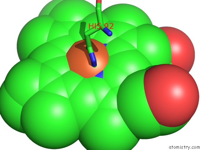
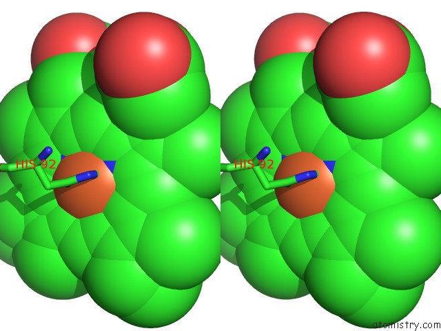
Iron binding site 3 out of 4 in 3gqg
Go back to
Iron binding site 3 out
of 4 in the Crystal Structure at Acidic pH of the Ferric Form of the Root Effect Hemoglobin From Trematomus Bernacchii.

Mono view

Stereo pair view
Mono view
Stereo pair view
A full contact list of Iron with other atoms in the Fe binding
site number 3 of Crystal Structure at Acidic pH of the Ferric Form of the Root Effect Hemoglobin From Trematomus Bernacchii. within 5.0Å range:
|
Iron binding site 4 out of 4 in 3gqg
Go back to
Iron binding site 4 out
of 4 in the Crystal Structure at Acidic pH of the Ferric Form of the Root Effect Hemoglobin From Trematomus Bernacchii.

Mono view
Stereo pair view
Mono view
Stereo pair view
A full contact list of Iron with other atoms in the Fe binding
site number 4 of Crystal Structure at Acidic pH of the Ferric Form of the Root Effect Hemoglobin From Trematomus Bernacchii. within 5.0Å range:
|
Reference:
A.Vergara,
M.Franzese,
A.Merlino,
G.Bonomi,
C.Verde,
D.Giordano,
G.Di Prisco,
H.C.Lee,
J.Peisach,
L.Mazzarella.
Correlation Between Hemichrome Stability and the Root Effect in Tetrameric Hemoglobins. Biophys.J. V. 97 866 2009.
ISSN: ISSN 0006-3495
PubMed: 19651045
DOI: 10.1016/J.BPJ.2009.04.056
Page generated: Sun Aug 4 10:51:19 2024
ISSN: ISSN 0006-3495
PubMed: 19651045
DOI: 10.1016/J.BPJ.2009.04.056
Last articles
F in 4TY9F in 4TXN
F in 4TVJ
F in 4TS2
F in 4TS0
F in 4TKG
F in 4TN4
F in 4TN6
F in 4S04
F in 4TLR