Atomistry » Iron » PDB 3gck-3h33 » 3gyx
Atomistry »
  Iron »
    PDB 3gck-3h33 »
      3gyx »

Iron in PDB 3gyx: Crystal Structure of Adenylylsulfate Reductase From Desulfovibrio Gigas

Protein crystallography data

The structure of Crystal Structure of Adenylylsulfate Reductase From Desulfovibrio Gigas, PDB code: 3gyx was solved by Y.-L.Chiang, Y.-C.Hsieh, E.-H.Liu, M.-Y.Liu, C.-J.Chen, with X-Ray Crystallography technique. A brief refinement statistics is given in the table below:

Resolution Low / High (Å) 30.00 / 3.20
Space group P 31 2 1
Cell size a, b, c (Å), α, β, γ (°) 199.629, 199.629, 317.422, 90.00, 90.00, 120.00
R / Rfree (%) 19.2 / 24.5

Iron Binding Sites:

Pages:

>>> Page 1 <<< Page 2, Binding sites: 11 - 20; Page 3, Binding sites: 21 - 30; Page 4, Binding sites: 31 - 40; Page 5, Binding sites: 41 - 48;

Binding sites:

The binding sites of Iron atom in the Crystal Structure of Adenylylsulfate Reductase From Desulfovibrio Gigas (pdb code 3gyx). This binding sites where shown within 5.0 Angstroms radius around Iron atom.
In total 48 binding sites of Iron where determined in the Crystal Structure of Adenylylsulfate Reductase From Desulfovibrio Gigas, PDB code: 3gyx:
Jump to Iron binding site number: 1; 2; 3; 4; 5; 6; 7; 8; 9; 10;

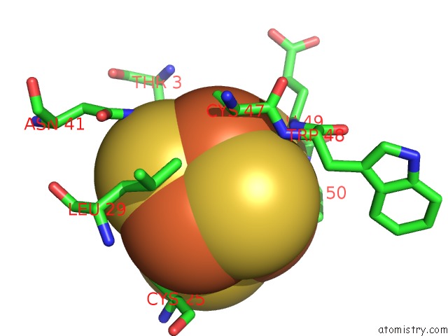
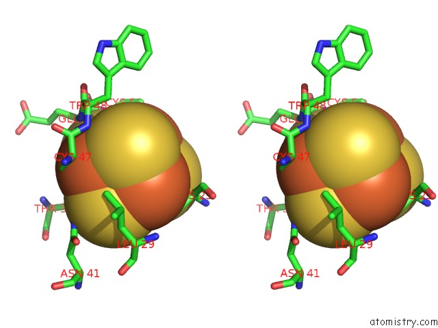
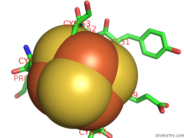
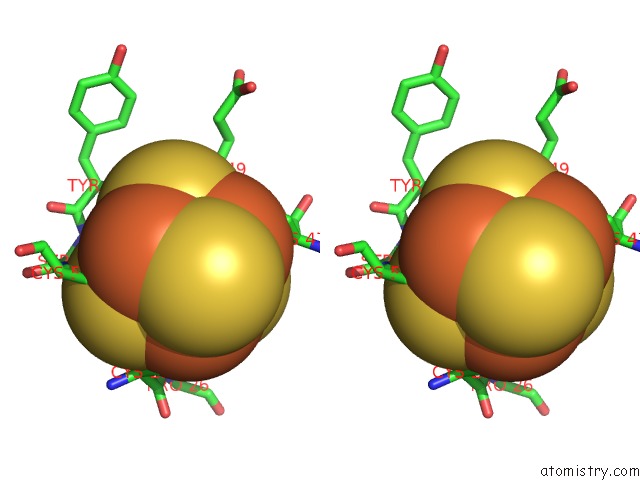
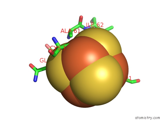
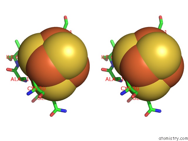
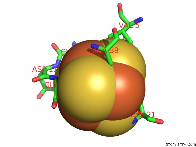
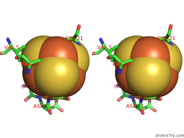
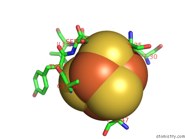
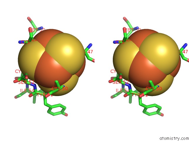
Iron binding site 1 out of 48 in 3gyx

Go back to Iron Binding Sites List in 3gyx
Iron binding site 1 out of 48 in the Crystal Structure of Adenylylsulfate Reductase From Desulfovibrio Gigas


Mono view


Stereo pair view

A full contact list of Iron with other atoms in the Fe binding site number 1 of Crystal Structure of Adenylylsulfate Reductase From Desulfovibrio Gigas within 5.0Å range:
probe atom residue distance (Å) B Occ
B:Fe1000

b:34.4
occ:1.00
FE1 B:SF41000 0.0 34.4 1.0
S3 B:SF41000 2.3 34.9 1.0
S2 B:SF41000 2.3 34.7 1.0
S4 B:SF41000 2.3 34.7 1.0
SG B:CYS47 2.3 33.6 1.0
FE4 B:SF41000 2.7 35.0 1.0
FE2 B:SF41000 2.7 33.8 1.0
FE3 B:SF41000 2.7 35.2 1.0
CB B:CYS47 3.5 33.5 1.0
CA B:CYS47 3.8 33.5 1.0
S1 B:SF41000 3.9 34.9 1.0
N B:GLU49 4.0 34.3 1.0
N B:TRP48 4.0 33.6 1.0
CD1 B:LEU29 4.0 38.0 1.0
C B:CYS47 4.3 33.5 1.0
OG1 B:THR3 4.4 41.8 1.0
CA B:GLU49 4.4 34.6 1.0
CG2 B:THR3 4.5 41.7 1.0
ND2 B:ASN41 4.5 37.1 1.0
N B:CYS50 4.6 34.7 1.0
SG B:CYS25 4.8 39.8 1.0
SG B:CYS50 4.8 34.5 1.0
C B:TRP48 4.9 34.1 1.0
SG B:CYS53 4.9 34.3 1.0

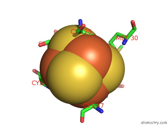
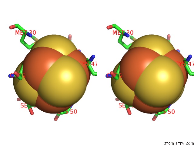
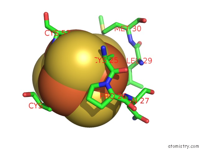
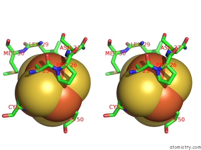
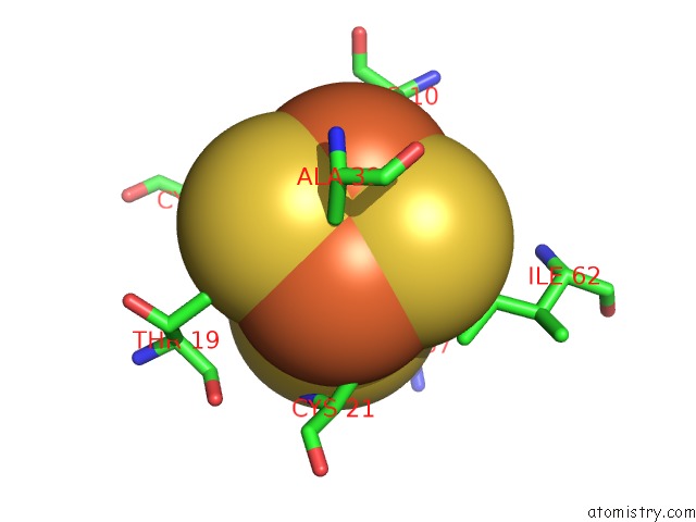
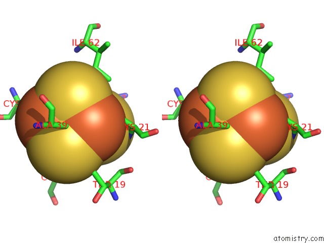
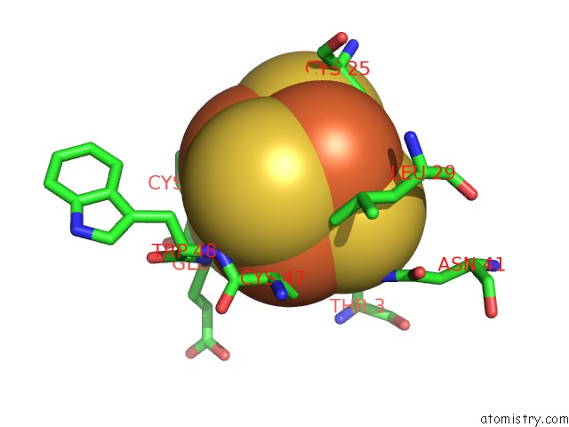
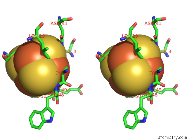
Iron binding site 2 out of 48 in 3gyx

Go back to Iron Binding Sites List in 3gyx
Iron binding site 2 out of 48 in the Crystal Structure of Adenylylsulfate Reductase From Desulfovibrio Gigas


Mono view


Stereo pair view

A full contact list of Iron with other atoms in the Fe binding site number 2 of Crystal Structure of Adenylylsulfate Reductase From Desulfovibrio Gigas within 5.0Å range:
probe atom residue distance (Å) B Occ
B:Fe1000

b:33.8
occ:1.00
FE2 B:SF41000 0.0 33.8 1.0
S1 B:SF41000 2.3 34.9 1.0
SG B:CYS53 2.3 34.3 1.0
S4 B:SF41000 2.3 34.7 1.0
S3 B:SF41000 2.3 34.9 1.0
FE3 B:SF41000 2.7 35.2 1.0
FE1 B:SF41000 2.7 34.4 1.0
FE4 B:SF41000 2.7 35.0 1.0
CB B:CYS53 3.5 34.7 1.0
OG1 B:THR3 3.9 41.8 1.0
S2 B:SF41000 3.9 34.7 1.0
CG2 B:THR3 3.9 41.7 1.0
N B:CYS53 4.1 34.9 1.0
CA B:CYS53 4.4 34.8 1.0
CB B:THR3 4.5 41.9 1.0
SG B:CYS47 4.5 33.6 1.0
CG B:MET30 4.6 38.6 1.0
SG B:CYS25 4.7 39.8 1.0
SD B:MET30 4.8 38.3 1.0
SG B:CYS50 4.9 34.5 1.0
N B:SER52 5.0 35.3 1.0

Iron binding site 3 out of 48 in 3gyx

Go back to Iron Binding Sites List in 3gyx
Iron binding site 3 out of 48 in the Crystal Structure of Adenylylsulfate Reductase From Desulfovibrio Gigas


Mono view


Stereo pair view

A full contact list of Iron with other atoms in the Fe binding site number 3 of Crystal Structure of Adenylylsulfate Reductase From Desulfovibrio Gigas within 5.0Å range:
probe atom residue distance (Å) B Occ
B:Fe1000

b:35.2
occ:1.00
FE3 B:SF41000 0.0 35.2 1.0
S1 B:SF41000 2.3 34.9 1.0
S4 B:SF41000 2.3 34.7 1.0
SG B:CYS50 2.3 34.5 1.0
S2 B:SF41000 2.3 34.7 1.0
FE4 B:SF41000 2.7 35.0 1.0
FE2 B:SF41000 2.7 33.8 1.0
FE1 B:SF41000 2.7 34.4 1.0
N B:CYS50 3.6 34.7 1.0
CB B:CYS50 3.7 34.8 1.0
S3 B:SF41000 3.9 34.9 1.0
N B:TYR51 4.0 35.2 1.0
CA B:CYS50 4.0 34.9 1.0
N B:SER52 4.1 35.3 1.0
CD B:PRO26 4.2 39.1 1.0
C B:CYS50 4.2 35.1 1.0
CB B:SER52 4.4 35.2 1.0
SG B:CYS53 4.6 34.3 1.0
OG B:SER52 4.7 35.4 1.0
C B:GLU49 4.7 34.6 1.0
N B:CYS53 4.7 34.9 1.0
N B:GLU49 4.7 34.3 1.0
SG B:CYS47 4.8 33.6 1.0
CA B:SER52 4.8 35.2 1.0
SG B:CYS25 4.8 39.8 1.0
CA B:TYR51 4.8 35.4 1.0
CA B:GLU49 4.9 34.6 1.0
C B:TYR51 4.9 35.4 1.0
CG B:PRO26 5.0 38.9 1.0

Iron binding site 4 out of 48 in 3gyx

Go back to Iron Binding Sites List in 3gyx
Iron binding site 4 out of 48 in the Crystal Structure of Adenylylsulfate Reductase From Desulfovibrio Gigas


Mono view


Stereo pair view

A full contact list of Iron with other atoms in the Fe binding site number 4 of Crystal Structure of Adenylylsulfate Reductase From Desulfovibrio Gigas within 5.0Å range:
probe atom residue distance (Å) B Occ
B:Fe1000

b:35.0
occ:1.00
FE4 B:SF41000 0.0 35.0 1.0
S3 B:SF41000 2.3 34.9 1.0
S2 B:SF41000 2.3 34.7 1.0
SG B:CYS25 2.3 39.8 1.0
S1 B:SF41000 2.3 34.9 1.0
FE3 B:SF41000 2.7 35.2 1.0
FE1 B:SF41000 2.7 34.4 1.0
FE2 B:SF41000 2.7 33.8 1.0
CB B:CYS25 3.4 39.9 1.0
S4 B:SF41000 3.9 34.7 1.0
CA B:CYS25 3.9 39.9 1.0
CD B:PRO26 4.0 39.1 1.0
CD1 B:LEU29 4.1 38.0 1.0
CB B:LEU29 4.5 38.3 1.0
CG B:LEU29 4.5 38.0 1.0
N B:PRO26 4.5 39.2 1.0
C B:CYS25 4.5 39.6 1.0
SG B:CYS50 4.6 34.5 1.0
SG B:CYS53 4.7 34.3 1.0
N B:ASN27 4.8 38.6 1.0
CG B:MET30 4.9 38.6 1.0
SG B:CYS47 4.9 33.6 1.0

Iron binding site 5 out of 48 in 3gyx

Go back to Iron Binding Sites List in 3gyx
Iron binding site 5 out of 48 in the Crystal Structure of Adenylylsulfate Reductase From Desulfovibrio Gigas


Mono view


Stereo pair view

A full contact list of Iron with other atoms in the Fe binding site number 5 of Crystal Structure of Adenylylsulfate Reductase From Desulfovibrio Gigas within 5.0Å range:
probe atom residue distance (Å) B Occ
B:Fe2000

b:50.8
occ:1.00
FE1 B:SF42000 0.0 50.8 1.0
S4 B:SF42000 2.3 50.8 1.0
S3 B:SF42000 2.3 50.9 1.0
S2 B:SF42000 2.3 50.8 1.0
SG B:CYS57 2.4 35.8 1.0
FE2 B:SF42000 2.7 52.0 1.0
FE3 B:SF42000 2.7 50.6 1.0
FE4 B:SF42000 2.7 49.2 1.0
CB B:CYS57 3.5 36.0 1.0
S1 B:SF42000 3.9 50.7 1.0
CA B:CYS57 4.2 36.1 1.0
CD B:PRO58 4.3 36.7 1.0
CB B:ALA61 4.4 39.1 1.0
CD1 B:ILE62 4.4 39.4 1.0
CG1 B:ILE62 4.5 39.3 1.0
SG B:CYS10 4.8 48.0 1.0
N B:PRO58 4.8 36.7 1.0
C B:CYS57 4.8 36.4 1.0
SG B:CYS21 4.8 45.5 1.0
N B:ALA61 4.8 38.9 1.0
CB B:CYS21 5.0 46.1 1.0
N B:GLN59 5.0 37.6 1.0
CB B:GLN59 5.0 37.9 1.0

Iron binding site 6 out of 48 in 3gyx

Go back to Iron Binding Sites List in 3gyx
Iron binding site 6 out of 48 in the Crystal Structure of Adenylylsulfate Reductase From Desulfovibrio Gigas


Mono view


Stereo pair view

A full contact list of Iron with other atoms in the Fe binding site number 6 of Crystal Structure of Adenylylsulfate Reductase From Desulfovibrio Gigas within 5.0Å range:
probe atom residue distance (Å) B Occ
B:Fe2000

b:52.0
occ:1.00
FE2 B:SF42000 0.0 52.0 1.0
S3 B:SF42000 2.3 50.9 1.0
S1 B:SF42000 2.3 50.7 1.0
S4 B:SF42000 2.3 50.8 1.0
FE1 B:SF42000 2.7 50.8 1.0
SG B:CYS13 2.7 51.1 1.0
FE3 B:SF42000 2.7 50.6 1.0
FE4 B:SF42000 2.7 49.2 1.0
N B:CYS13 3.7 51.5 1.0
S2 B:SF42000 3.9 50.8 1.0
CB B:CYS13 4.0 51.9 1.0
C B:GLY12 4.1 51.1 1.0
CA B:CYS13 4.3 51.9 1.0
CA B:GLY12 4.3 50.6 1.0
N B:GLY12 4.4 50.2 1.0
OD2 B:ASP11 4.5 50.7 1.0
CD B:PRO58 4.6 36.7 1.0
SG B:CYS21 4.7 45.5 1.0
SG B:CYS10 4.7 48.0 1.0
CG2 B:THR19 4.8 50.1 1.0
SG B:CYS57 4.8 35.8 1.0
O B:GLY12 4.9 51.1 1.0
N B:ALA20 4.9 48.8 1.0

Iron binding site 7 out of 48 in 3gyx

Go back to Iron Binding Sites List in 3gyx
Iron binding site 7 out of 48 in the Crystal Structure of Adenylylsulfate Reductase From Desulfovibrio Gigas


Mono view


Stereo pair view

A full contact list of Iron with other atoms in the Fe binding site number 7 of Crystal Structure of Adenylylsulfate Reductase From Desulfovibrio Gigas within 5.0Å range:
probe atom residue distance (Å) B Occ
B:Fe2000

b:50.6
occ:1.00
FE3 B:SF42000 0.0 50.6 1.0
S4 B:SF42000 2.3 50.8 1.0
S2 B:SF42000 2.3 50.8 1.0
S1 B:SF42000 2.3 50.7 1.0
SG B:CYS21 2.3 45.5 1.0
FE2 B:SF42000 2.7 52.0 1.0
FE1 B:SF42000 2.7 50.8 1.0
FE4 B:SF42000 2.7 49.2 1.0
CB B:CYS21 3.2 46.1 1.0
CB B:ALA39 3.8 40.5 1.0
S3 B:SF42000 3.9 50.9 1.0
N B:CYS21 4.1 46.9 1.0
CA B:CYS21 4.3 46.1 1.0
CG2 B:THR19 4.4 50.1 1.0
CD1 B:ILE62 4.5 39.4 1.0
SG B:CYS13 4.8 51.1 1.0
SG B:CYS10 4.8 48.0 1.0
SG B:CYS57 4.9 35.8 1.0

Iron binding site 8 out of 48 in 3gyx

Go back to Iron Binding Sites List in 3gyx
Iron binding site 8 out of 48 in the Crystal Structure of Adenylylsulfate Reductase From Desulfovibrio Gigas


Mono view


Stereo pair view

A full contact list of Iron with other atoms in the Fe binding site number 8 of Crystal Structure of Adenylylsulfate Reductase From Desulfovibrio Gigas within 5.0Å range:
probe atom residue distance (Å) B Occ
B:Fe2000

b:49.2
occ:1.00
FE4 B:SF42000 0.0 49.2 1.0
SG B:CYS10 2.3 48.0 1.0
S1 B:SF42000 2.3 50.7 1.0
S2 B:SF42000 2.3 50.8 1.0
S3 B:SF42000 2.3 50.9 1.0
FE2 B:SF42000 2.7 52.0 1.0
FE3 B:SF42000 2.7 50.6 1.0
FE1 B:SF42000 2.7 50.8 1.0
CB B:CYS10 3.3 48.1 1.0
S4 B:SF42000 3.9 50.8 1.0
CA B:CYS10 4.0 48.2 1.0
N B:GLY12 4.0 50.2 1.0
CB B:ALA39 4.1 40.5 1.0
CA B:GLY12 4.3 50.6 1.0
N B:ASP11 4.3 49.0 1.0
CG1 B:VAL5 4.5 42.9 1.0
N B:ALA39 4.5 41.0 1.0
C B:CYS10 4.5 48.6 1.0
CB B:ALA61 4.8 39.1 1.0
SG B:CYS21 4.9 45.5 1.0
SG B:CYS57 4.9 35.8 1.0
CA B:ALA39 5.0 40.5 1.0

Iron binding site 9 out of 48 in 3gyx

Go back to Iron Binding Sites List in 3gyx
Iron binding site 9 out of 48 in the Crystal Structure of Adenylylsulfate Reductase From Desulfovibrio Gigas


Mono view


Stereo pair view

A full contact list of Iron with other atoms in the Fe binding site number 9 of Crystal Structure of Adenylylsulfate Reductase From Desulfovibrio Gigas within 5.0Å range:
probe atom residue distance (Å) B Occ
D:Fe1000

b:30.5
occ:1.00
FE1 D:SF41000 0.0 30.5 1.0
SG D:CYS47 2.3 31.1 1.0
S3 D:SF41000 2.3 31.9 1.0
S4 D:SF41000 2.3 31.6 1.0
S2 D:SF41000 2.3 31.9 1.0
FE2 D:SF41000 2.7 30.6 1.0
FE3 D:SF41000 2.7 32.0 1.0
FE4 D:SF41000 2.7 30.7 1.0
CB D:CYS47 3.5 31.2 1.0
N D:GLU49 3.8 31.6 1.0
CA D:CYS47 3.9 31.3 1.0
S1 D:SF41000 3.9 31.3 1.0
N D:TRP48 4.0 31.7 1.0
CD1 D:LEU29 4.1 36.8 1.0
CG2 D:THR3 4.2 39.3 1.0
CA D:GLU49 4.2 31.5 1.0
C D:CYS47 4.3 31.5 1.0
OG1 D:THR3 4.3 39.4 1.0
N D:CYS50 4.5 31.3 1.0
ND2 D:ASN41 4.6 34.0 1.0
SG D:CYS53 4.8 31.2 1.0
SG D:CYS50 4.8 31.2 1.0
C D:TRP48 4.9 31.7 1.0
CB D:THR3 4.9 39.5 1.0
C D:GLU49 4.9 31.5 1.0
SG D:CYS25 4.9 37.2 1.0
CA D:TRP48 5.0 31.9 1.0

Iron binding site 10 out of 48 in 3gyx

Go back to Iron Binding Sites List in 3gyx
Iron binding site 10 out of 48 in the Crystal Structure of Adenylylsulfate Reductase From Desulfovibrio Gigas


Mono view


Stereo pair view

A full contact list of Iron with other atoms in the Fe binding site number 10 of Crystal Structure of Adenylylsulfate Reductase From Desulfovibrio Gigas within 5.0Å range:
probe atom residue distance (Å) B Occ
D:Fe1000

b:30.6
occ:1.00
FE2 D:SF41000 0.0 30.6 1.0
SG D:CYS53 2.3 31.2 1.0
S3 D:SF41000 2.3 31.9 1.0
S4 D:SF41000 2.3 31.6 1.0
S1 D:SF41000 2.3 31.3 1.0
FE3 D:SF41000 2.7 32.0 1.0
FE4 D:SF41000 2.7 30.7 1.0
FE1 D:SF41000 2.7 30.5 1.0
CB D:CYS53 3.4 31.3 1.0
CG2 D:THR3 3.8 39.3 1.0
N D:CYS53 3.9 31.4 1.0
S2 D:SF41000 3.9 31.9 1.0
OG1 D:THR3 4.1 39.4 1.0
CA D:CYS53 4.3 31.3 1.0
CB D:THR3 4.5 39.5 1.0
N D:SER52 4.6 31.3 1.0
SG D:CYS47 4.7 31.1 1.0
SG D:CYS50 4.7 31.2 1.0
SG D:CYS25 4.7 37.2 1.0
CG D:MET30 4.8 37.5 1.0
N D:TYR51 4.8 31.2 1.0
N D:ILE54 4.9 31.3 1.0
C D:SER52 5.0 31.4 1.0

Reference:

Y.-L.Chiang, Y.-C.Hsieh, J.-Y.Fang, E.-H.Liu, Y.-C.Huang, P.Chuankhayan, J.Jeyakanthan, M.-Y.Liu, S.I.Chan, C.-J.Chen. Crystal Structure of Adenylylsulfate Reductase From Desulfovibrio Gigas Suggests A Potential Self-Regulation Mechanism Involving the C Terminus of the Beta-Subunit J.Bacteriol. V. 191 7597 2009.
ISSN: ISSN 0021-9193
PubMed: 19820092
DOI: 10.1128/JB.00583-09
Page generated: Sun Aug 4 10:59:16 2024

Last articles

Zn in 9J0N
Zn in 9J0O
Zn in 9J0P
Zn in 9FJX
Zn in 9EKB
Zn in 9C0F
Zn in 9CAH
Zn in 9CH0
Zn in 9CH3
Zn in 9CH1
© Copyright 2008-2020 by atomistry.com
Home   |    Site Map   |    Copyright   |    Contact us   |    Privacy