Iron »
PDB 3gck-3h33 »
3h31 »
Iron in PDB 3h31: Structure of Rhodothermus Marinus Hipip at 1.0 A Resolution
Protein crystallography data
The structure of Structure of Rhodothermus Marinus Hipip at 1.0 A Resolution, PDB code: 3h31
was solved by
M.Stelter,
A.M.P.Melo,
L.Saraiva,
M.Teixeira,
M.Archer,
with X-Ray Crystallography technique. A brief refinement statistics is given in the table below:
| Resolution Low / High (Å) | 10.00 / 1.00 |
| Space group | P 21 21 21 |
| Cell size a, b, c (Å), α, β, γ (°) | 27.267, 43.343, 46.787, 90.00, 90.00, 90.00 |
| R / Rfree (%) | 10.4 / 14.4 |
Iron Binding Sites:
The binding sites of Iron atom in the Structure of Rhodothermus Marinus Hipip at 1.0 A Resolution
(pdb code 3h31). This binding sites where shown within
5.0 Angstroms radius around Iron atom.
In total 4 binding sites of Iron where determined in the Structure of Rhodothermus Marinus Hipip at 1.0 A Resolution, PDB code: 3h31:
Jump to Iron binding site number: 1; 2; 3; 4;
In total 4 binding sites of Iron where determined in the Structure of Rhodothermus Marinus Hipip at 1.0 A Resolution, PDB code: 3h31:
Jump to Iron binding site number: 1; 2; 3; 4;
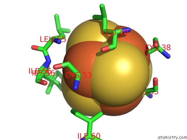
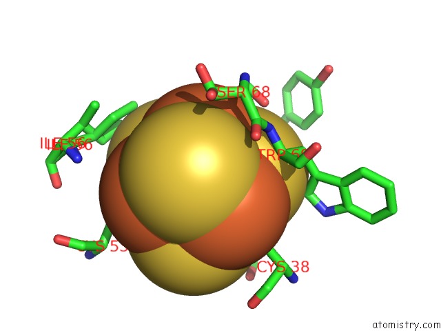
Iron binding site 1 out of 4 in 3h31
Go back to
Iron binding site 1 out
of 4 in the Structure of Rhodothermus Marinus Hipip at 1.0 A Resolution
Mono view
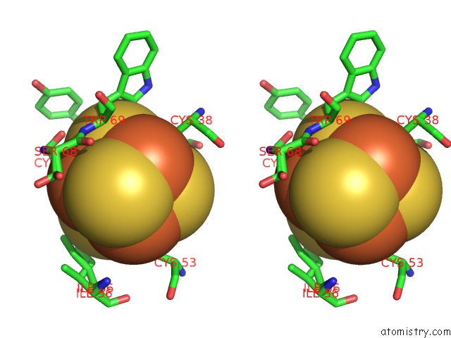
Stereo pair view
Mono view
Stereo pair view
A full contact list of Iron with other atoms in the Fe binding
site number 1 of Structure of Rhodothermus Marinus Hipip at 1.0 A Resolution within 5.0Å range:
|
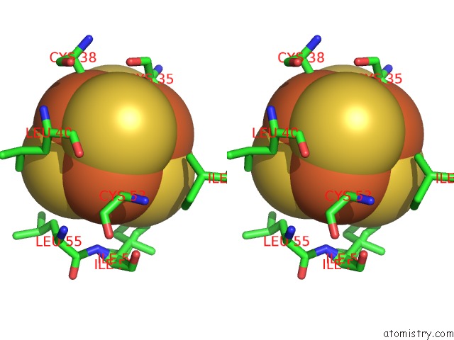
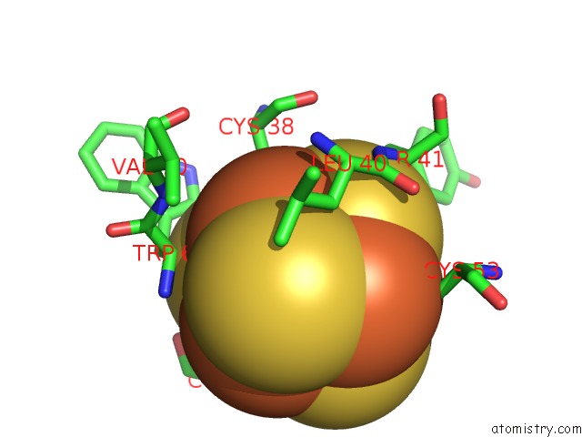
Iron binding site 2 out of 4 in 3h31
Go back to
Iron binding site 2 out
of 4 in the Structure of Rhodothermus Marinus Hipip at 1.0 A Resolution
Mono view
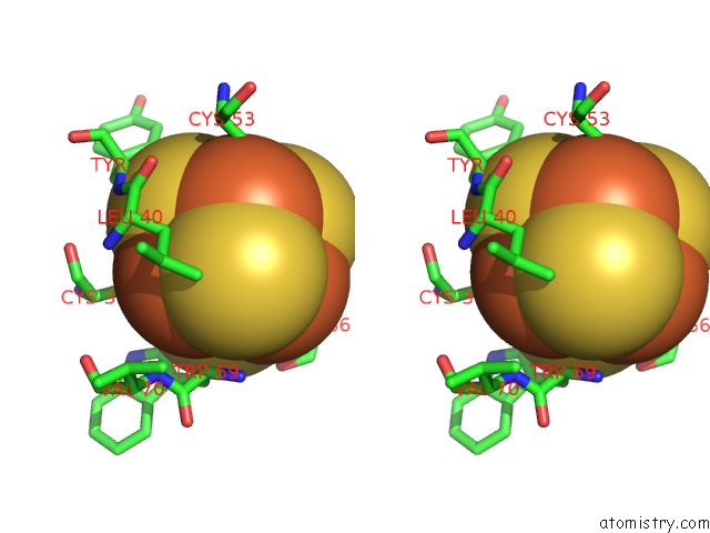
Stereo pair view
Mono view
Stereo pair view
A full contact list of Iron with other atoms in the Fe binding
site number 2 of Structure of Rhodothermus Marinus Hipip at 1.0 A Resolution within 5.0Å range:
|
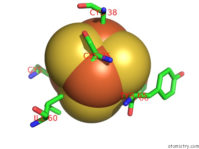
Iron binding site 3 out of 4 in 3h31
Go back to
Iron binding site 3 out
of 4 in the Structure of Rhodothermus Marinus Hipip at 1.0 A Resolution
Mono view
Stereo pair view
Mono view
Stereo pair view
A full contact list of Iron with other atoms in the Fe binding
site number 3 of Structure of Rhodothermus Marinus Hipip at 1.0 A Resolution within 5.0Å range:
|
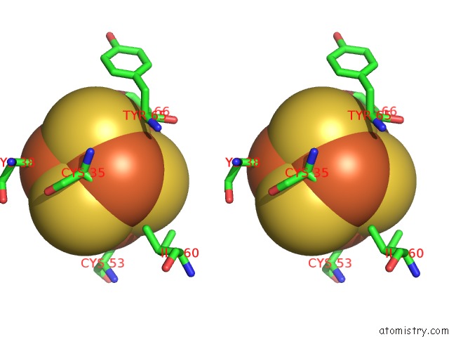
Iron binding site 4 out of 4 in 3h31
Go back to
Iron binding site 4 out
of 4 in the Structure of Rhodothermus Marinus Hipip at 1.0 A Resolution
Mono view
Stereo pair view
Mono view
Stereo pair view
A full contact list of Iron with other atoms in the Fe binding
site number 4 of Structure of Rhodothermus Marinus Hipip at 1.0 A Resolution within 5.0Å range:
|
Reference:
M.Stelter,
A.M.P.Melo,
G.O.Hreggvidsson,
S.Hjorleifsdottir,
L.M.Saraiva,
M.Teixeira,
M.Archer.
Structure at 1.0 A Resolution of A High-Potential Iron-Sulfur Protein Involved in the Aerobic Respiratory Chain of Rhodothermus Marinus J.Biol.Inorg.Chem. V. 15 303 2010.
ISSN: ISSN 0949-8257
PubMed: 20225399
DOI: 10.1007/S00775-009-0603-8
Page generated: Sun Aug 4 11:04:13 2024
ISSN: ISSN 0949-8257
PubMed: 20225399
DOI: 10.1007/S00775-009-0603-8
Last articles
Cl in 5XYOCl in 5XXI
Cl in 5XXK
Cl in 5XWE
Cl in 5XVU
Cl in 5XVG
Cl in 5XVF
Cl in 5XVA
Cl in 5XT8
Cl in 5XUH