Iron »
PDB 3h34-3hni »
3h58 »
Iron in PDB 3h58: Myoglobin Cavity Mutant H64LV68N Met Form
Protein crystallography data
The structure of Myoglobin Cavity Mutant H64LV68N Met Form, PDB code: 3h58
was solved by
J.Soman,
J.S.Olson,
with X-Ray Crystallography technique. A brief refinement statistics is given in the table below:
| Resolution Low / High (Å) | 39.03 / 1.80 |
| Space group | P 6 |
| Cell size a, b, c (Å), α, β, γ (°) | 90.130, 90.130, 45.260, 90.00, 90.00, 120.00 |
| R / Rfree (%) | 19.6 / 21.9 |
Iron Binding Sites:
The binding sites of Iron atom in the Myoglobin Cavity Mutant H64LV68N Met Form
(pdb code 3h58). This binding sites where shown within
5.0 Angstroms radius around Iron atom.
In total only one binding site of Iron was determined in the Myoglobin Cavity Mutant H64LV68N Met Form, PDB code: 3h58:
In total only one binding site of Iron was determined in the Myoglobin Cavity Mutant H64LV68N Met Form, PDB code: 3h58:
Iron binding site 1 out of 1 in 3h58
Go back to
Iron binding site 1 out
of 1 in the Myoglobin Cavity Mutant H64LV68N Met Form
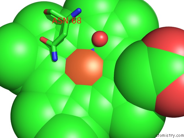
Mono view
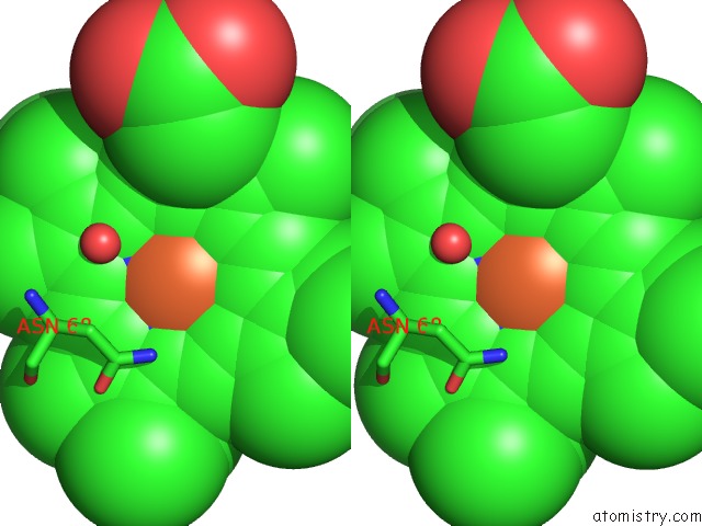
Stereo pair view
Mono view
Stereo pair view
A full contact list of Iron with other atoms in the Fe binding
site number 1 of Myoglobin Cavity Mutant H64LV68N Met Form within 5.0Å range:
|
Reference:
R.A.Goldbeck,
M.L.Pillsbury,
R.A.Jensen,
J.L.Mendoza,
R.L.Nguyen,
J.S.Olson,
J.Soman,
D.S.Kliger,
R.M.Esquerra.
Optical Detection of Disordered Water Within A Protein Cavity. J.Am.Chem.Soc. V. 131 12265 2009.
ISSN: ISSN 0002-7863
PubMed: 19655795
DOI: 10.1021/JA903409J
Page generated: Sun Aug 4 11:23:31 2024
ISSN: ISSN 0002-7863
PubMed: 19655795
DOI: 10.1021/JA903409J
Last articles
Fe in 2YXOFe in 2YRS
Fe in 2YXC
Fe in 2YNM
Fe in 2YVJ
Fe in 2YP1
Fe in 2YU2
Fe in 2YU1
Fe in 2YQB
Fe in 2YOO