Iron »
PDB 3h34-3hni »
3h65 »
Iron in PDB 3h65: The Crystal Structure of C176A Mutated [Fe]-Hydrogenase (Hmd) Holoenzyme in Complex with Methylenetetrahydromethanopterin
Enzymatic activity of The Crystal Structure of C176A Mutated [Fe]-Hydrogenase (Hmd) Holoenzyme in Complex with Methylenetetrahydromethanopterin
All present enzymatic activity of The Crystal Structure of C176A Mutated [Fe]-Hydrogenase (Hmd) Holoenzyme in Complex with Methylenetetrahydromethanopterin:
1.12.98.2;
1.12.98.2;
Protein crystallography data
The structure of The Crystal Structure of C176A Mutated [Fe]-Hydrogenase (Hmd) Holoenzyme in Complex with Methylenetetrahydromethanopterin, PDB code: 3h65
was solved by
T.Hiromoto,
E.Warkentin,
S.Shima,
U.Ermler,
with X-Ray Crystallography technique. A brief refinement statistics is given in the table below:
| Resolution Low / High (Å) | 48.81 / 2.15 |
| Space group | I 41 2 2 |
| Cell size a, b, c (Å), α, β, γ (°) | 97.630, 97.630, 165.750, 90.00, 90.00, 90.00 |
| R / Rfree (%) | 17 / 20.3 |
Iron Binding Sites:
The binding sites of Iron atom in the The Crystal Structure of C176A Mutated [Fe]-Hydrogenase (Hmd) Holoenzyme in Complex with Methylenetetrahydromethanopterin
(pdb code 3h65). This binding sites where shown within
5.0 Angstroms radius around Iron atom.
In total only one binding site of Iron was determined in the The Crystal Structure of C176A Mutated [Fe]-Hydrogenase (Hmd) Holoenzyme in Complex with Methylenetetrahydromethanopterin, PDB code: 3h65:
In total only one binding site of Iron was determined in the The Crystal Structure of C176A Mutated [Fe]-Hydrogenase (Hmd) Holoenzyme in Complex with Methylenetetrahydromethanopterin, PDB code: 3h65:
Iron binding site 1 out of 1 in 3h65
Go back to
Iron binding site 1 out
of 1 in the The Crystal Structure of C176A Mutated [Fe]-Hydrogenase (Hmd) Holoenzyme in Complex with Methylenetetrahydromethanopterin
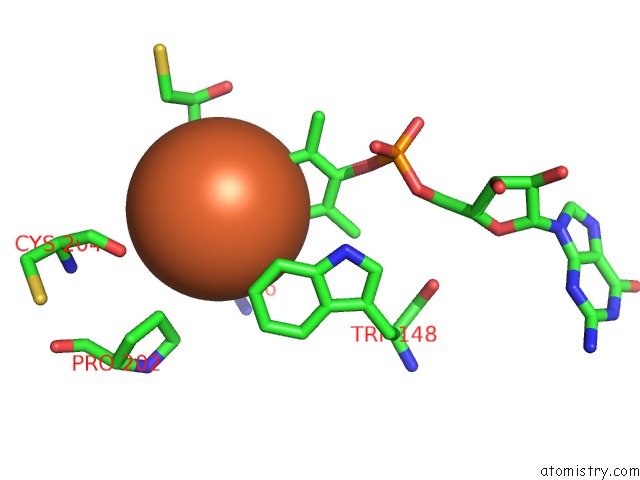
Mono view
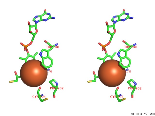
Stereo pair view
Mono view
Stereo pair view
A full contact list of Iron with other atoms in the Fe binding
site number 1 of The Crystal Structure of C176A Mutated [Fe]-Hydrogenase (Hmd) Holoenzyme in Complex with Methylenetetrahydromethanopterin within 5.0Å range:
|
Reference:
T.Hiromoto,
E.Warkentin,
J.Moll,
U.Ermler,
S.Shima.
The Crystal Structure of An [Fe]-Hydrogenase-Substrate Complex Reveals the Framework For H2 Activation. Angew.Chem.Int.Ed.Engl. V. 48 6457 2009.
ISSN: ISSN 1433-7851
PubMed: 19623593
DOI: 10.1002/ANIE.200902695
Page generated: Sun Aug 4 11:23:31 2024
ISSN: ISSN 1433-7851
PubMed: 19623593
DOI: 10.1002/ANIE.200902695
Last articles
Zn in 9MJ5Zn in 9HNW
Zn in 9G0L
Zn in 9FNE
Zn in 9DZN
Zn in 9E0I
Zn in 9D32
Zn in 9DAK
Zn in 8ZXC
Zn in 8ZUF