Iron »
PDB 3h34-3hni »
3h7j »
Iron in PDB 3h7j: Crystal Structure of Bacb, An Enzyme Involved in Bacilysin Synthesis, in Monoclinic Form
Protein crystallography data
The structure of Crystal Structure of Bacb, An Enzyme Involved in Bacilysin Synthesis, in Monoclinic Form, PDB code: 3h7j
was solved by
M.Rajavel,
B.Gopal,
with X-Ray Crystallography technique. A brief refinement statistics is given in the table below:
| Resolution Low / High (Å) | 26.21 / 1.87 |
| Space group | P 1 21 1 |
| Cell size a, b, c (Å), α, β, γ (°) | 45.990, 118.493, 46.926, 90.00, 97.92, 90.00 |
| R / Rfree (%) | 18.5 / 22.7 |
Other elements in 3h7j:
The structure of Crystal Structure of Bacb, An Enzyme Involved in Bacilysin Synthesis, in Monoclinic Form also contains other interesting chemical elements:
| Cobalt | (Co) | 4 atoms |
Iron Binding Sites:
The binding sites of Iron atom in the Crystal Structure of Bacb, An Enzyme Involved in Bacilysin Synthesis, in Monoclinic Form
(pdb code 3h7j). This binding sites where shown within
5.0 Angstroms radius around Iron atom.
In total 2 binding sites of Iron where determined in the Crystal Structure of Bacb, An Enzyme Involved in Bacilysin Synthesis, in Monoclinic Form, PDB code: 3h7j:
Jump to Iron binding site number: 1; 2;
In total 2 binding sites of Iron where determined in the Crystal Structure of Bacb, An Enzyme Involved in Bacilysin Synthesis, in Monoclinic Form, PDB code: 3h7j:
Jump to Iron binding site number: 1; 2;
Iron binding site 1 out of 2 in 3h7j
Go back to
Iron binding site 1 out
of 2 in the Crystal Structure of Bacb, An Enzyme Involved in Bacilysin Synthesis, in Monoclinic Form
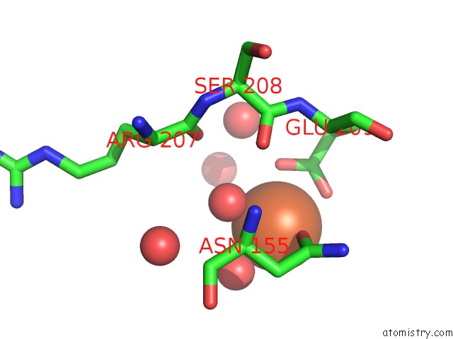
Mono view
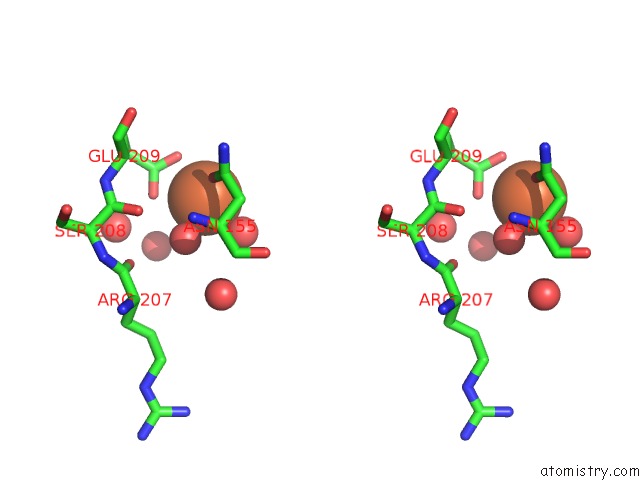
Stereo pair view
Mono view
Stereo pair view
A full contact list of Iron with other atoms in the Fe binding
site number 1 of Crystal Structure of Bacb, An Enzyme Involved in Bacilysin Synthesis, in Monoclinic Form within 5.0Å range:
|
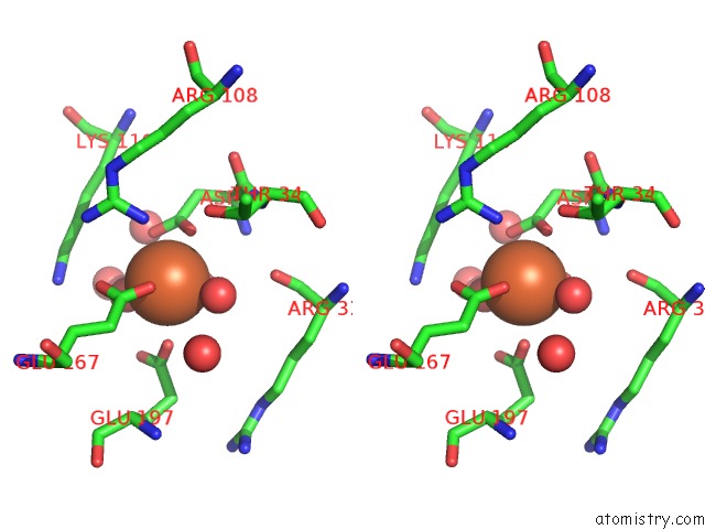
Iron binding site 2 out of 2 in 3h7j
Go back to
Iron binding site 2 out
of 2 in the Crystal Structure of Bacb, An Enzyme Involved in Bacilysin Synthesis, in Monoclinic Form
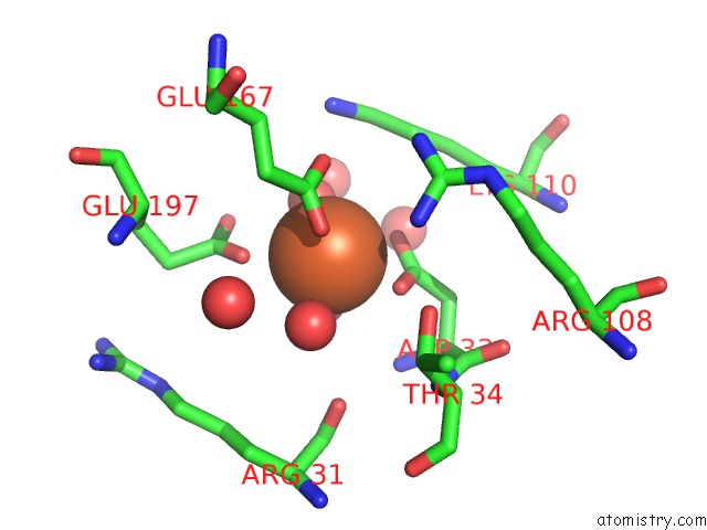
Mono view
Stereo pair view
Mono view
Stereo pair view
A full contact list of Iron with other atoms in the Fe binding
site number 2 of Crystal Structure of Bacb, An Enzyme Involved in Bacilysin Synthesis, in Monoclinic Form within 5.0Å range:
|
Reference:
M.Rajavel,
A.Mitra,
B.Gopal.
Role of Bacillus Subtilis Bacb in the Synthesis of Bacilysin J.Biol.Chem. V. 284 31882 2009.
ISSN: ISSN 0021-9258
PubMed: 19776011
DOI: 10.1074/JBC.M109.014522
Page generated: Sun Aug 4 11:23:31 2024
ISSN: ISSN 0021-9258
PubMed: 19776011
DOI: 10.1074/JBC.M109.014522
Last articles
Zn in 9MJ5Zn in 9HNW
Zn in 9G0L
Zn in 9FNE
Zn in 9DZN
Zn in 9E0I
Zn in 9D32
Zn in 9DAK
Zn in 8ZXC
Zn in 8ZUF