Iron »
PDB 3h34-3hni »
3hb3 »
Iron in PDB 3hb3: High Resolution Crystal Structure of Paracoccus Denitrificans Cytochrome C Oxidase
Enzymatic activity of High Resolution Crystal Structure of Paracoccus Denitrificans Cytochrome C Oxidase
All present enzymatic activity of High Resolution Crystal Structure of Paracoccus Denitrificans Cytochrome C Oxidase:
1.9.3.1;
1.9.3.1;
Protein crystallography data
The structure of High Resolution Crystal Structure of Paracoccus Denitrificans Cytochrome C Oxidase, PDB code: 3hb3
was solved by
J.Koepke,
H.Angerer,
G.Peng,
with X-Ray Crystallography technique. A brief refinement statistics is given in the table below:
| Resolution Low / High (Å) | 19.98 / 2.25 |
| Space group | P 21 21 21 |
| Cell size a, b, c (Å), α, β, γ (°) | 83.403, 150.473, 157.185, 90.00, 90.00, 90.00 |
| R / Rfree (%) | 21.8 / 28 |
Other elements in 3hb3:
The structure of High Resolution Crystal Structure of Paracoccus Denitrificans Cytochrome C Oxidase also contains other interesting chemical elements:
| Manganese | (Mn) | 1 atom |
| Copper | (Cu) | 3 atoms |
| Calcium | (Ca) | 1 atom |
Iron Binding Sites:
The binding sites of Iron atom in the High Resolution Crystal Structure of Paracoccus Denitrificans Cytochrome C Oxidase
(pdb code 3hb3). This binding sites where shown within
5.0 Angstroms radius around Iron atom.
In total 2 binding sites of Iron where determined in the High Resolution Crystal Structure of Paracoccus Denitrificans Cytochrome C Oxidase, PDB code: 3hb3:
Jump to Iron binding site number: 1; 2;
In total 2 binding sites of Iron where determined in the High Resolution Crystal Structure of Paracoccus Denitrificans Cytochrome C Oxidase, PDB code: 3hb3:
Jump to Iron binding site number: 1; 2;
Iron binding site 1 out of 2 in 3hb3
Go back to
Iron binding site 1 out
of 2 in the High Resolution Crystal Structure of Paracoccus Denitrificans Cytochrome C Oxidase
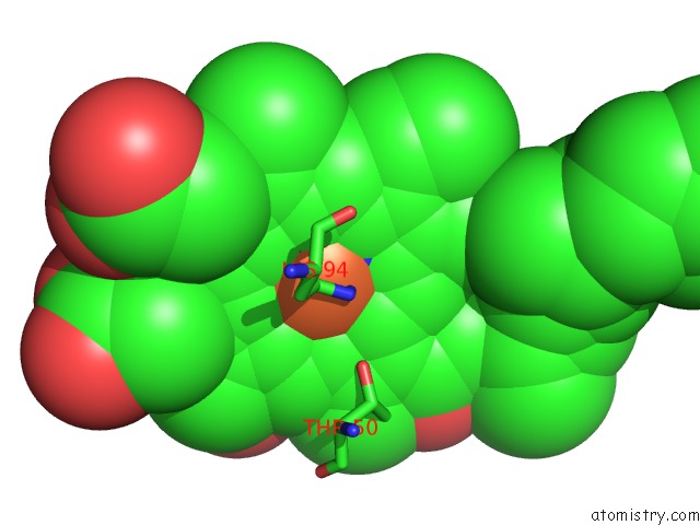
Mono view
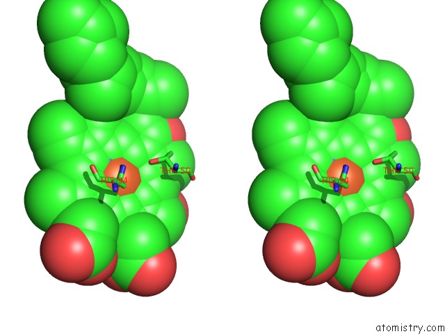
Stereo pair view
Mono view
Stereo pair view
A full contact list of Iron with other atoms in the Fe binding
site number 1 of High Resolution Crystal Structure of Paracoccus Denitrificans Cytochrome C Oxidase within 5.0Å range:
|
Iron binding site 2 out of 2 in 3hb3
Go back to
Iron binding site 2 out
of 2 in the High Resolution Crystal Structure of Paracoccus Denitrificans Cytochrome C Oxidase
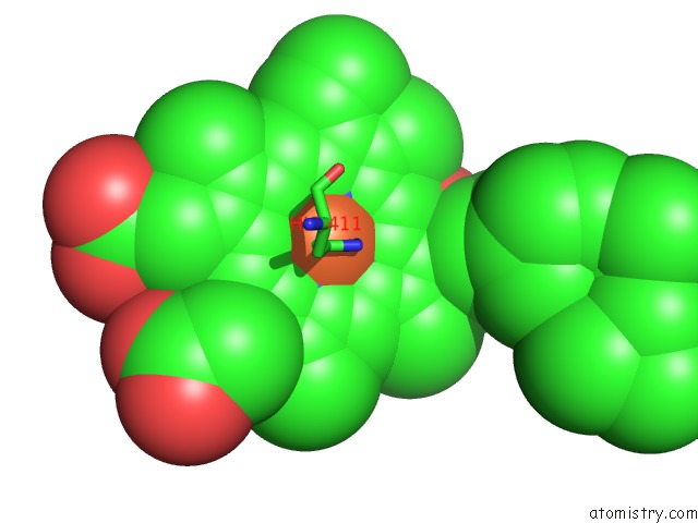
Mono view

Stereo pair view
Mono view
Stereo pair view
A full contact list of Iron with other atoms in the Fe binding
site number 2 of High Resolution Crystal Structure of Paracoccus Denitrificans Cytochrome C Oxidase within 5.0Å range:
|
Reference:
J.Koepke,
E.Olkhova,
H.Angerer,
H.Muller,
G.Peng,
H.Michel.
High Resolution Crystal Structure of Paracoccus Denitrificans Cytochrome C Oxidase: New Insights Into the Active Site and the Proton Transfer Pathways Biochim.Biophys.Acta V.1787 635 2009.
ISSN: ISSN 0006-3002
PubMed: 19374884
DOI: 10.1016/J.BBABIO.2009.04.003
Page generated: Sun Aug 4 11:24:39 2024
ISSN: ISSN 0006-3002
PubMed: 19374884
DOI: 10.1016/J.BBABIO.2009.04.003
Last articles
Zn in 9J0NZn in 9J0O
Zn in 9J0P
Zn in 9FJX
Zn in 9EKB
Zn in 9C0F
Zn in 9CAH
Zn in 9CH0
Zn in 9CH3
Zn in 9CH1