Iron »
PDB 3h34-3hni »
3hdl »
Iron in PDB 3hdl: Crystal Structure of Highly Glycosylated Peroxidase From Royal Palm Tree
Protein crystallography data
The structure of Crystal Structure of Highly Glycosylated Peroxidase From Royal Palm Tree, PDB code: 3hdl
was solved by
L.Watanabe,
P.R.Moura,
L.Bleicher,
A.S.Nascimento,
L.S.Zamorano,
J.J.Calvete,
S.Bursakov,
M.G.Roig,
V.L.Shnyrov,
I.Polikarpov,
with X-Ray Crystallography technique. A brief refinement statistics is given in the table below:
| Resolution Low / High (Å) | 44.78 / 1.85 |
| Space group | P 31 2 1 |
| Cell size a, b, c (Å), α, β, γ (°) | 117.821, 117.821, 93.447, 90.00, 90.00, 120.00 |
| R / Rfree (%) | 17.6 / 18.7 |
Other elements in 3hdl:
The structure of Crystal Structure of Highly Glycosylated Peroxidase From Royal Palm Tree also contains other interesting chemical elements:
| Calcium | (Ca) | 2 atoms |
Iron Binding Sites:
The binding sites of Iron atom in the Crystal Structure of Highly Glycosylated Peroxidase From Royal Palm Tree
(pdb code 3hdl). This binding sites where shown within
5.0 Angstroms radius around Iron atom.
In total only one binding site of Iron was determined in the Crystal Structure of Highly Glycosylated Peroxidase From Royal Palm Tree, PDB code: 3hdl:
In total only one binding site of Iron was determined in the Crystal Structure of Highly Glycosylated Peroxidase From Royal Palm Tree, PDB code: 3hdl:
Iron binding site 1 out of 1 in 3hdl
Go back to
Iron binding site 1 out
of 1 in the Crystal Structure of Highly Glycosylated Peroxidase From Royal Palm Tree
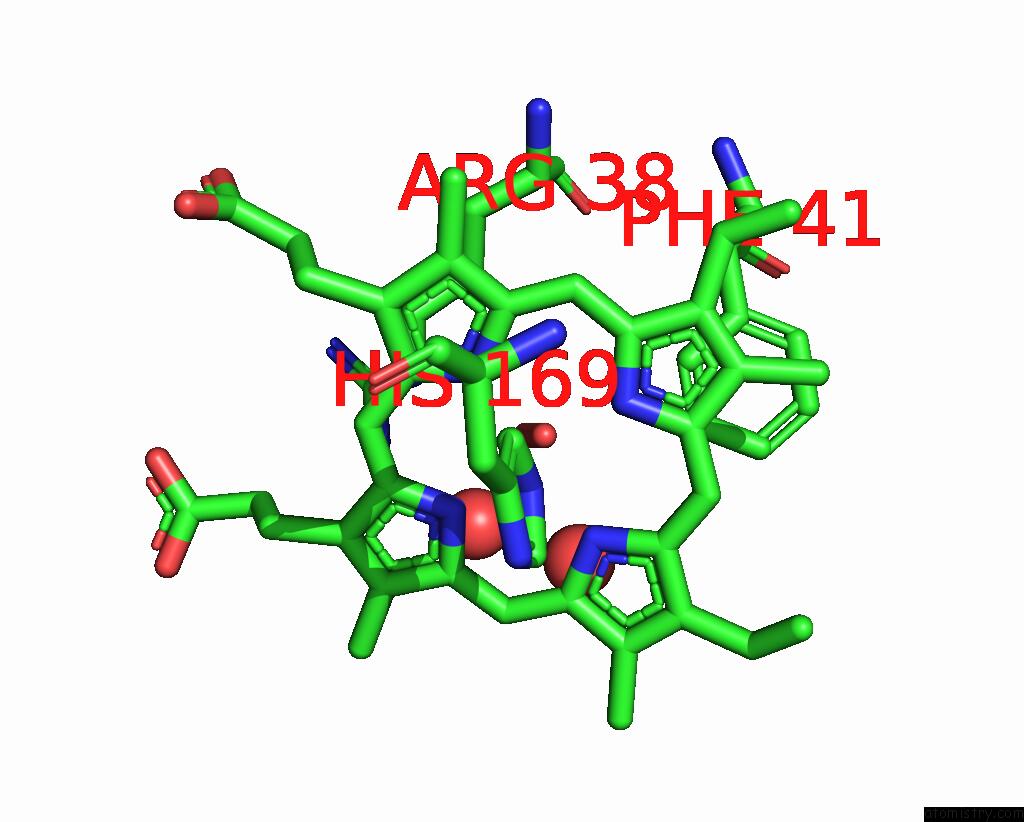
Mono view
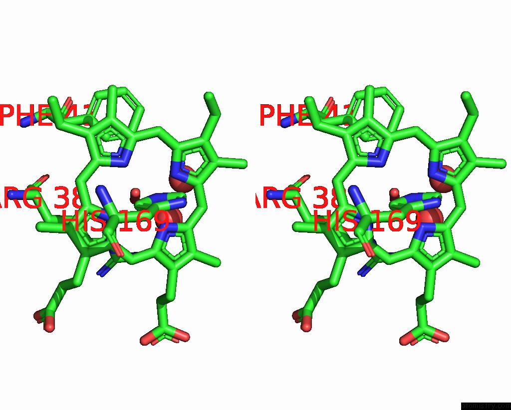
Stereo pair view
Mono view
Stereo pair view
A full contact list of Iron with other atoms in the Fe binding
site number 1 of Crystal Structure of Highly Glycosylated Peroxidase From Royal Palm Tree within 5.0Å range:
|
Reference:
L.Watanabe,
P.R.De Moura,
L.Bleicher,
A.S.Nascimento,
L.S.Zamorano,
J.J.Calvete,
L.Sanz,
A.Perez,
S.Bursakov,
M.G.Roig,
V.L.Shnyrov,
I.Polikarpov.
Crystal Structure and Statistical Coupling Analysis of Highly Glycosylated Peroxidase From Royal Palm Tree (Roystonea Regia). J.Struct.Biol. V. 169 226 2010.
ISSN: ISSN 1047-8477
PubMed: 19854274
DOI: 10.1016/J.JSB.2009.10.009
Page generated: Sun Aug 4 11:26:11 2024
ISSN: ISSN 1047-8477
PubMed: 19854274
DOI: 10.1016/J.JSB.2009.10.009
Last articles
Zn in 9J0NZn in 9J0O
Zn in 9J0P
Zn in 9FJX
Zn in 9EKB
Zn in 9C0F
Zn in 9CAH
Zn in 9CH0
Zn in 9CH3
Zn in 9CH1