Iron »
PDB 3h34-3hni »
3hfb »
Iron in PDB 3hfb: Crystal Structure of Human Tryoptophan Hydroxylase Type 1 with Lp- 534193
Enzymatic activity of Crystal Structure of Human Tryoptophan Hydroxylase Type 1 with Lp- 534193
All present enzymatic activity of Crystal Structure of Human Tryoptophan Hydroxylase Type 1 with Lp- 534193:
1.14.16.4;
1.14.16.4;
Protein crystallography data
The structure of Crystal Structure of Human Tryoptophan Hydroxylase Type 1 with Lp- 534193, PDB code: 3hfb
was solved by
L.W.Tari,
R.V.Swanson,
M.J.Hunter,
with X-Ray Crystallography technique. A brief refinement statistics is given in the table below:
| Resolution Low / High (Å) | N/A / 1.92 |
| Space group | P 1 21 1 |
| Cell size a, b, c (Å), α, β, γ (°) | 47.354, 58.143, 56.625, 90.00, 96.77, 90.00 |
| R / Rfree (%) | 21.4 / 28.9 |
Iron Binding Sites:
The binding sites of Iron atom in the Crystal Structure of Human Tryoptophan Hydroxylase Type 1 with Lp- 534193
(pdb code 3hfb). This binding sites where shown within
5.0 Angstroms radius around Iron atom.
In total only one binding site of Iron was determined in the Crystal Structure of Human Tryoptophan Hydroxylase Type 1 with Lp- 534193, PDB code: 3hfb:
In total only one binding site of Iron was determined in the Crystal Structure of Human Tryoptophan Hydroxylase Type 1 with Lp- 534193, PDB code: 3hfb:
Iron binding site 1 out of 1 in 3hfb
Go back to
Iron binding site 1 out
of 1 in the Crystal Structure of Human Tryoptophan Hydroxylase Type 1 with Lp- 534193
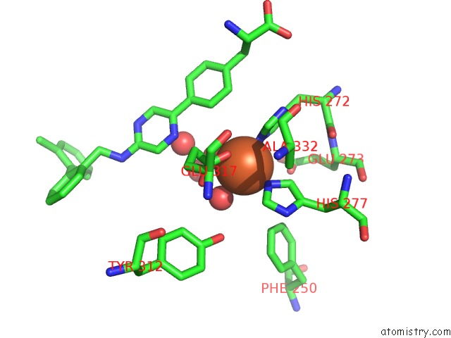
Mono view
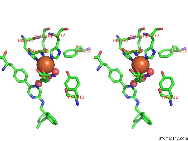
Stereo pair view
Mono view
Stereo pair view
A full contact list of Iron with other atoms in the Fe binding
site number 1 of Crystal Structure of Human Tryoptophan Hydroxylase Type 1 with Lp- 534193 within 5.0Å range:
|
Reference:
G.Cianchetta,
T.Stouch,
W.Yu,
Z.C.Shi,
L.W.Tari,
R.V.Swanson,
M.J.Hunter,
I.D.Hoffman,
Q.Liu.
Mechanism of Inhibition of Novel Tryptophan Hydroxylase Inhibitors Revealed By Co-Crystal Structures and Kinetic Analysis. Curr Chem Genomics V. 4 19 2010.
ISSN: ESSN 1875-3973
PubMed: 20556201
DOI: 10.2174/1875397301004010019
Page generated: Sun Aug 4 11:29:29 2024
ISSN: ESSN 1875-3973
PubMed: 20556201
DOI: 10.2174/1875397301004010019
Last articles
Zn in 9MJ5Zn in 9HNW
Zn in 9G0L
Zn in 9FNE
Zn in 9DZN
Zn in 9E0I
Zn in 9D32
Zn in 9DAK
Zn in 8ZXC
Zn in 8ZUF