Iron »
PDB 3hnj-3i8r »
3hsp »
Iron in PDB 3hsp: Ternary Structure of Neuronal Nitric Oxide Synthase with Nha and No Bound(2)
Enzymatic activity of Ternary Structure of Neuronal Nitric Oxide Synthase with Nha and No Bound(2)
All present enzymatic activity of Ternary Structure of Neuronal Nitric Oxide Synthase with Nha and No Bound(2):
1.14.13.39;
1.14.13.39;
Protein crystallography data
The structure of Ternary Structure of Neuronal Nitric Oxide Synthase with Nha and No Bound(2), PDB code: 3hsp
was solved by
T.Doukov,
H.Li,
M.Soltis,
T.L.Poulos,
with X-Ray Crystallography technique. A brief refinement statistics is given in the table below:
| Resolution Low / High (Å) | 19.82 / 2.20 |
| Space group | P 21 21 21 |
| Cell size a, b, c (Å), α, β, γ (°) | 51.820, 110.560, 164.530, 90.00, 90.00, 90.00 |
| R / Rfree (%) | 16.4 / 21.7 |
Other elements in 3hsp:
The structure of Ternary Structure of Neuronal Nitric Oxide Synthase with Nha and No Bound(2) also contains other interesting chemical elements:
| Zinc | (Zn) | 1 atom |
Iron Binding Sites:
The binding sites of Iron atom in the Ternary Structure of Neuronal Nitric Oxide Synthase with Nha and No Bound(2)
(pdb code 3hsp). This binding sites where shown within
5.0 Angstroms radius around Iron atom.
In total 2 binding sites of Iron where determined in the Ternary Structure of Neuronal Nitric Oxide Synthase with Nha and No Bound(2), PDB code: 3hsp:
Jump to Iron binding site number: 1; 2;
In total 2 binding sites of Iron where determined in the Ternary Structure of Neuronal Nitric Oxide Synthase with Nha and No Bound(2), PDB code: 3hsp:
Jump to Iron binding site number: 1; 2;
Iron binding site 1 out of 2 in 3hsp
Go back to
Iron binding site 1 out
of 2 in the Ternary Structure of Neuronal Nitric Oxide Synthase with Nha and No Bound(2)
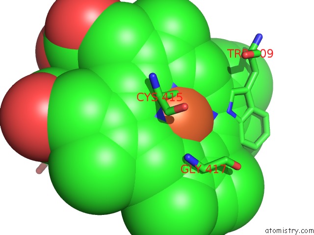
Mono view
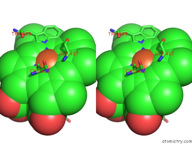
Stereo pair view
Mono view
Stereo pair view
A full contact list of Iron with other atoms in the Fe binding
site number 1 of Ternary Structure of Neuronal Nitric Oxide Synthase with Nha and No Bound(2) within 5.0Å range:
|
Iron binding site 2 out of 2 in 3hsp
Go back to
Iron binding site 2 out
of 2 in the Ternary Structure of Neuronal Nitric Oxide Synthase with Nha and No Bound(2)
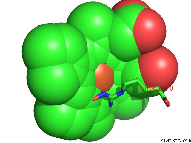
Mono view
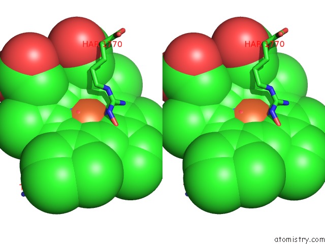
Stereo pair view
Mono view
Stereo pair view
A full contact list of Iron with other atoms in the Fe binding
site number 2 of Ternary Structure of Neuronal Nitric Oxide Synthase with Nha and No Bound(2) within 5.0Å range:
|
Reference:
T.Doukov,
H.Li,
M.Soltis,
T.L.Poulos.
Single Crystal Structural and Absorption Spectral Characterizations of Nitric Oxide Synthase Complexed with N(Omega)-Hydroxy-L-Arginine and Diatomic Ligands. Biochemistry V. 48 10246 2009.
ISSN: ISSN 0006-2960
PubMed: 19791770
DOI: 10.1021/BI9009743
Page generated: Sun Aug 4 11:46:48 2024
ISSN: ISSN 0006-2960
PubMed: 19791770
DOI: 10.1021/BI9009743
Last articles
Zn in 9MJ5Zn in 9HNW
Zn in 9G0L
Zn in 9FNE
Zn in 9DZN
Zn in 9E0I
Zn in 9D32
Zn in 9DAK
Zn in 8ZXC
Zn in 8ZUF