Iron »
PDB 3i9t-3j7b »
3i9t »
Iron in PDB 3i9t: Crystal Structure of the Rat Heme Oxygenase (Ho-1) in Complex with Heme Binding Dithiothreitol (Dtt)
Enzymatic activity of Crystal Structure of the Rat Heme Oxygenase (Ho-1) in Complex with Heme Binding Dithiothreitol (Dtt)
All present enzymatic activity of Crystal Structure of the Rat Heme Oxygenase (Ho-1) in Complex with Heme Binding Dithiothreitol (Dtt):
1.14.99.3;
1.14.99.3;
Protein crystallography data
The structure of Crystal Structure of the Rat Heme Oxygenase (Ho-1) in Complex with Heme Binding Dithiothreitol (Dtt), PDB code: 3i9t
was solved by
T.Matsui,
M.Unno,
M.Ikeda-Saito,
with X-Ray Crystallography technique. A brief refinement statistics is given in the table below:
| Resolution Low / High (Å) | 30.00 / 2.15 |
| Space group | P 32 2 1 |
| Cell size a, b, c (Å), α, β, γ (°) | 65.739, 65.739, 120.663, 90.00, 90.00, 120.00 |
| R / Rfree (%) | 16.2 / 20.9 |
Iron Binding Sites:
The binding sites of Iron atom in the Crystal Structure of the Rat Heme Oxygenase (Ho-1) in Complex with Heme Binding Dithiothreitol (Dtt)
(pdb code 3i9t). This binding sites where shown within
5.0 Angstroms radius around Iron atom.
In total only one binding site of Iron was determined in the Crystal Structure of the Rat Heme Oxygenase (Ho-1) in Complex with Heme Binding Dithiothreitol (Dtt), PDB code: 3i9t:
In total only one binding site of Iron was determined in the Crystal Structure of the Rat Heme Oxygenase (Ho-1) in Complex with Heme Binding Dithiothreitol (Dtt), PDB code: 3i9t:
Iron binding site 1 out of 1 in 3i9t
Go back to
Iron binding site 1 out
of 1 in the Crystal Structure of the Rat Heme Oxygenase (Ho-1) in Complex with Heme Binding Dithiothreitol (Dtt)
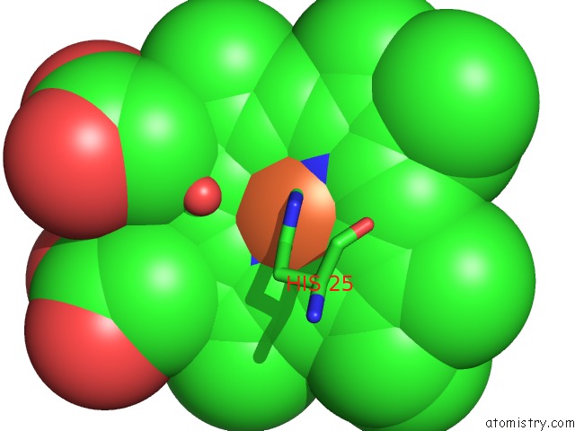
Mono view
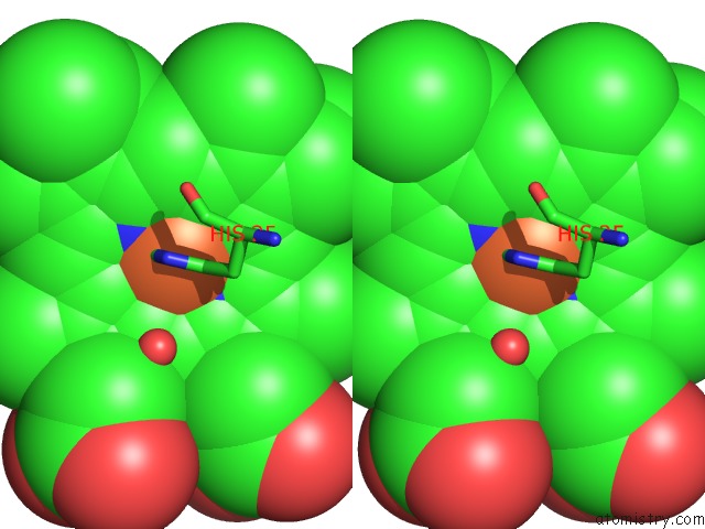
Stereo pair view
Mono view
Stereo pair view
A full contact list of Iron with other atoms in the Fe binding
site number 1 of Crystal Structure of the Rat Heme Oxygenase (Ho-1) in Complex with Heme Binding Dithiothreitol (Dtt) within 5.0Å range:
|
Reference:
T.Matsui,
M.Iwasaki,
R.Sugiyama,
M.Unno,
M.Ikeda-Saito.
Dioxygen Activation For the Self-Degradation of Heme: Reaction Mechanism and Regulation of Heme Oxygenase. Inorg.Chem. V. 49 3602 2010.
ISSN: ISSN 0020-1669
PubMed: 20380462
DOI: 10.1021/IC901869T
Page generated: Sun Aug 4 12:15:23 2024
ISSN: ISSN 0020-1669
PubMed: 20380462
DOI: 10.1021/IC901869T
Last articles
F in 4QJMF in 4QJ0
F in 4QJC
F in 4QJ5
F in 4QJ4
F in 4QAC
F in 4QJ3
F in 4QAB
F in 4QIY
F in 4QIZ