Iron »
PDB 3i9t-3j7b »
3ivy »
Iron in PDB 3ivy: Crystal Structure of Mycobacterium Tuberculosis Cytochrome P450 CYP125, P212121 Crystal Form
Protein crystallography data
The structure of Crystal Structure of Mycobacterium Tuberculosis Cytochrome P450 CYP125, P212121 Crystal Form, PDB code: 3ivy
was solved by
K.J.Mclean,
C.Levy,
A.W.Munro,
D.Leys,
with X-Ray Crystallography technique. A brief refinement statistics is given in the table below:
| Resolution Low / High (Å) | 28.22 / 1.35 |
| Space group | P 21 21 21 |
| Cell size a, b, c (Å), α, β, γ (°) | 60.030, 86.020, 89.630, 90.00, 90.00, 90.00 |
| R / Rfree (%) | 15.9 / 20.4 |
Iron Binding Sites:
The binding sites of Iron atom in the Crystal Structure of Mycobacterium Tuberculosis Cytochrome P450 CYP125, P212121 Crystal Form
(pdb code 3ivy). This binding sites where shown within
5.0 Angstroms radius around Iron atom.
In total only one binding site of Iron was determined in the Crystal Structure of Mycobacterium Tuberculosis Cytochrome P450 CYP125, P212121 Crystal Form, PDB code: 3ivy:
In total only one binding site of Iron was determined in the Crystal Structure of Mycobacterium Tuberculosis Cytochrome P450 CYP125, P212121 Crystal Form, PDB code: 3ivy:
Iron binding site 1 out of 1 in 3ivy
Go back to
Iron binding site 1 out
of 1 in the Crystal Structure of Mycobacterium Tuberculosis Cytochrome P450 CYP125, P212121 Crystal Form
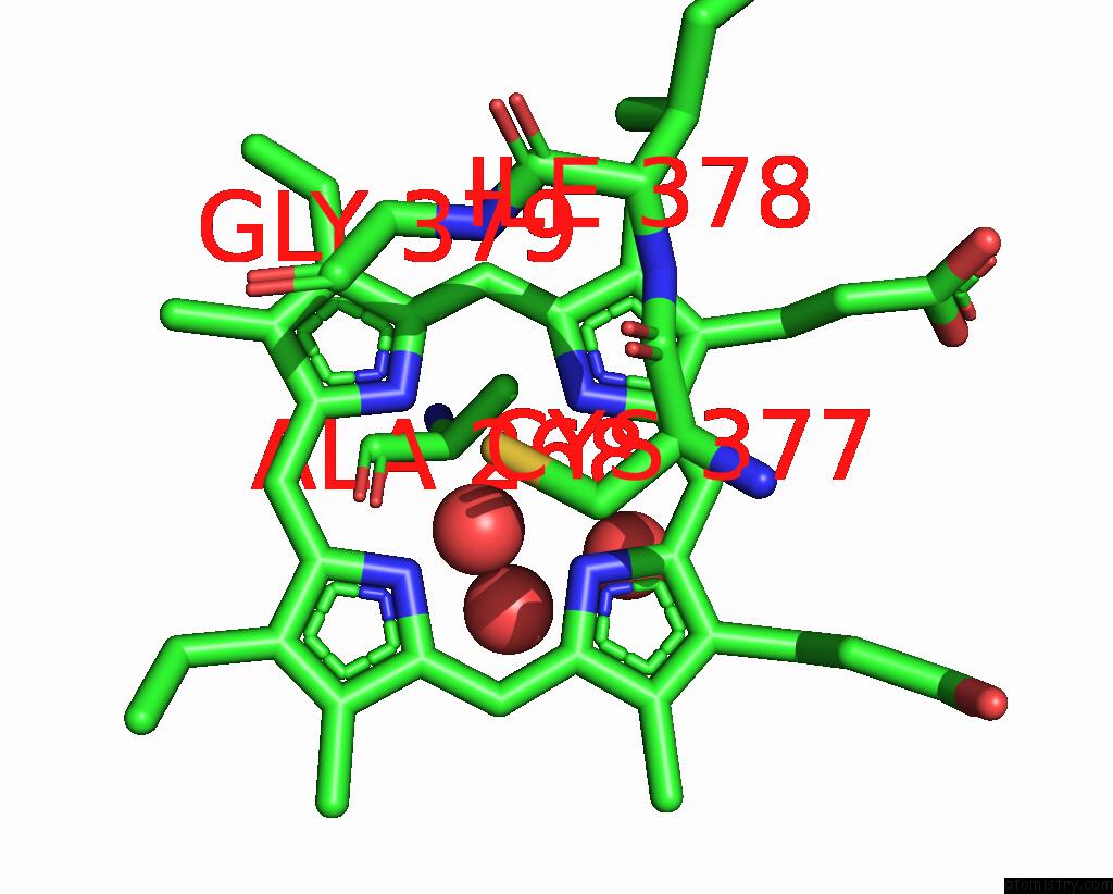
Mono view
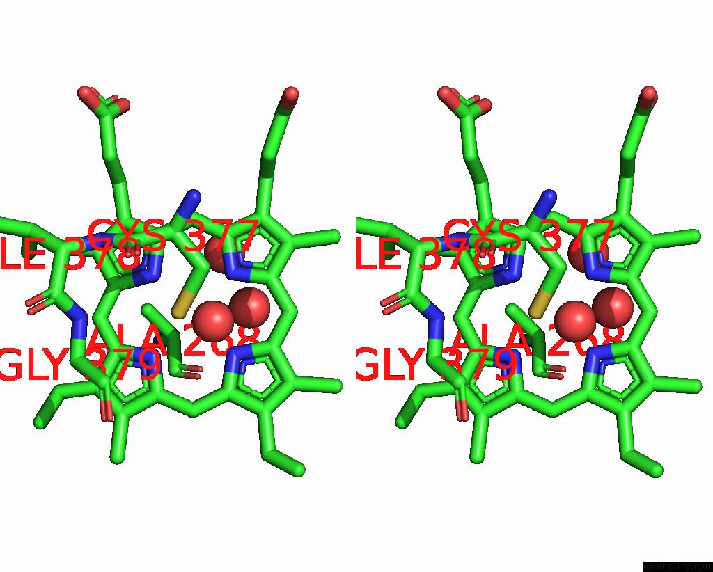
Stereo pair view
Mono view
Stereo pair view
A full contact list of Iron with other atoms in the Fe binding
site number 1 of Crystal Structure of Mycobacterium Tuberculosis Cytochrome P450 CYP125, P212121 Crystal Form within 5.0Å range:
|
Reference:
K.J.Mclean,
P.Lafite,
C.Levy,
M.R.Cheesman,
N.Mast,
I.A.Pikuleva,
D.Leys,
A.W.Munro.
The Structure of Mycobacterium Tuberculosis CYP125: Molecular Basis For Cholesterol Binding in A P450 Needed For Host Infection. J.Biol.Chem. V. 284 35524 2009.
ISSN: ISSN 0021-9258
PubMed: 19846552
DOI: 10.1074/JBC.M109.032706
Page generated: Sun Aug 4 12:35:02 2024
ISSN: ISSN 0021-9258
PubMed: 19846552
DOI: 10.1074/JBC.M109.032706
Last articles
Zn in 9J0NZn in 9J0O
Zn in 9J0P
Zn in 9FJX
Zn in 9EKB
Zn in 9C0F
Zn in 9CAH
Zn in 9CH0
Zn in 9CH3
Zn in 9CH1