Iron »
PDB 3k9z-3kyw »
3kcy »
Iron in PDB 3kcy: Factor Inhibiting Hif-1 Alpha in Complex with 8-Hydroxyquinoline
Enzymatic activity of Factor Inhibiting Hif-1 Alpha in Complex with 8-Hydroxyquinoline
All present enzymatic activity of Factor Inhibiting Hif-1 Alpha in Complex with 8-Hydroxyquinoline:
1.14.11.16;
1.14.11.16;
Protein crystallography data
The structure of Factor Inhibiting Hif-1 Alpha in Complex with 8-Hydroxyquinoline, PDB code: 3kcy
was solved by
H.Moon,
S.Han,
J.Choe,
with X-Ray Crystallography technique. A brief refinement statistics is given in the table below:
| Resolution Low / High (Å) | 42.14 / 2.59 |
| Space group | P 41 21 2 |
| Cell size a, b, c (Å), α, β, γ (°) | 86.912, 86.912, 144.559, 90.00, 90.00, 90.00 |
| R / Rfree (%) | 25.5 / 33.5 |
Iron Binding Sites:
The binding sites of Iron atom in the Factor Inhibiting Hif-1 Alpha in Complex with 8-Hydroxyquinoline
(pdb code 3kcy). This binding sites where shown within
5.0 Angstroms radius around Iron atom.
In total only one binding site of Iron was determined in the Factor Inhibiting Hif-1 Alpha in Complex with 8-Hydroxyquinoline, PDB code: 3kcy:
In total only one binding site of Iron was determined in the Factor Inhibiting Hif-1 Alpha in Complex with 8-Hydroxyquinoline, PDB code: 3kcy:
Iron binding site 1 out of 1 in 3kcy
Go back to
Iron binding site 1 out
of 1 in the Factor Inhibiting Hif-1 Alpha in Complex with 8-Hydroxyquinoline
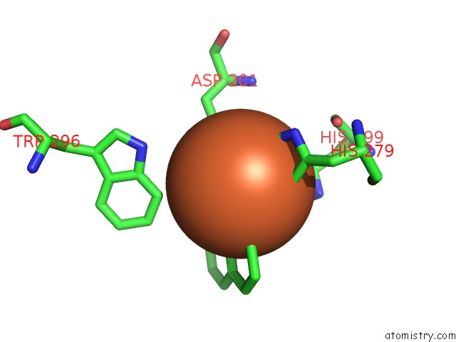
Mono view
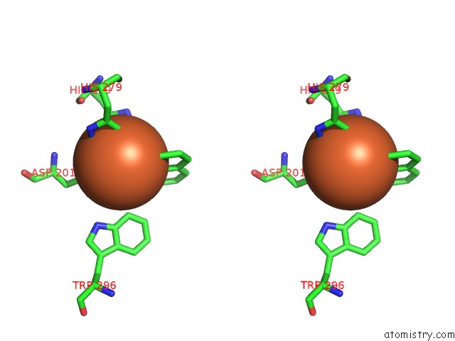
Stereo pair view
Mono view
Stereo pair view
A full contact list of Iron with other atoms in the Fe binding
site number 1 of Factor Inhibiting Hif-1 Alpha in Complex with 8-Hydroxyquinoline within 5.0Å range:
|
Reference:
H.Moon,
S.Han,
H.Park,
J.Choe.
Crystal Structures of Human Fih-1 in Complex with Quinol Family Inhibitors Mol.Cells V. 29 471 2010.
ISSN: ISSN 1016-8478
PubMed: 20396966
DOI: 10.1007/S10059-010-0058-3
Page generated: Sun Aug 4 13:54:23 2024
ISSN: ISSN 1016-8478
PubMed: 20396966
DOI: 10.1007/S10059-010-0058-3
Last articles
Fe in 2YXOFe in 2YRS
Fe in 2YXC
Fe in 2YNM
Fe in 2YVJ
Fe in 2YP1
Fe in 2YU2
Fe in 2YU1
Fe in 2YQB
Fe in 2YOO