Iron »
PDB 3k9z-3kyw »
3kun »
Iron in PDB 3kun: X-Ray Structure of the Metcyano Form of Dehaloperoxidase From Amphitrite Ornata: Evidence For Photoreductive Lysis of Iron-Cyanide Bond
Protein crystallography data
The structure of X-Ray Structure of the Metcyano Form of Dehaloperoxidase From Amphitrite Ornata: Evidence For Photoreductive Lysis of Iron-Cyanide Bond, PDB code: 3kun
was solved by
V.S.Serrano,
Z.Chen,
J.F.Gaff,
R.Rose,
S.Franzen,
with X-Ray Crystallography technique. A brief refinement statistics is given in the table below:
| Resolution Low / High (Å) | 35.00 / 1.26 |
| Space group | P 21 21 21 |
| Cell size a, b, c (Å), α, β, γ (°) | 57.252, 66.550, 68.713, 90.00, 90.00, 90.00 |
| R / Rfree (%) | 15.3 / 18.6 |
Iron Binding Sites:
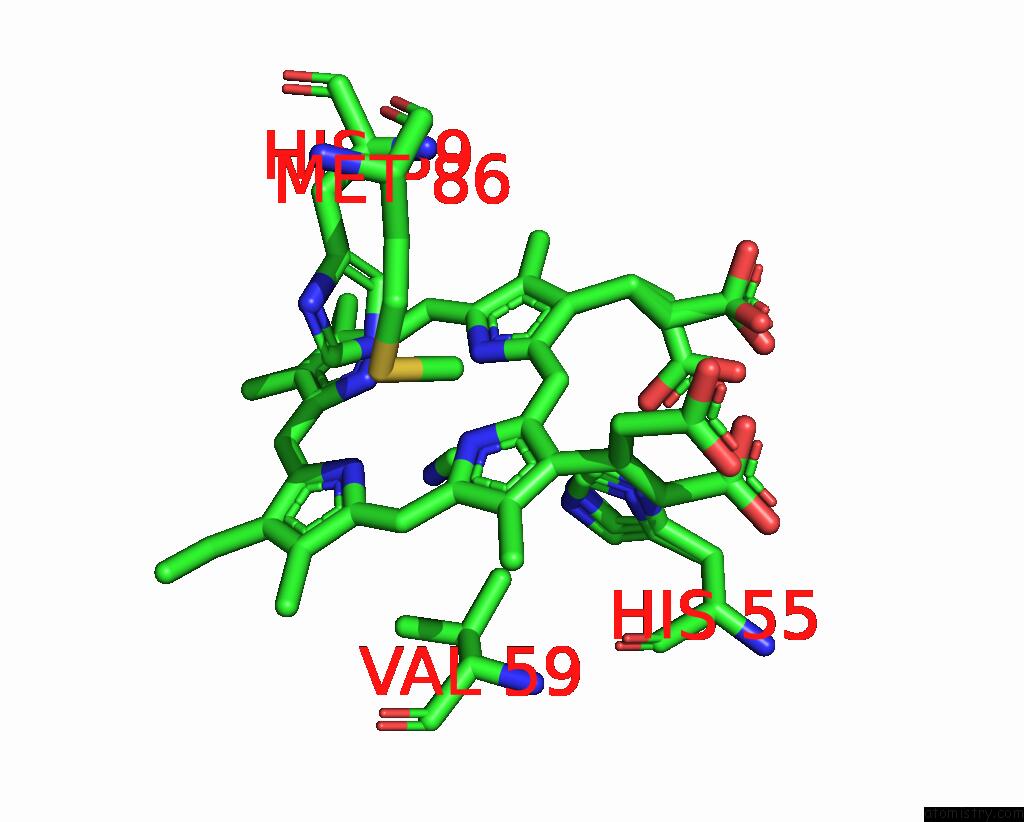
The binding sites of Iron atom in the X-Ray Structure of the Metcyano Form of Dehaloperoxidase From Amphitrite Ornata: Evidence For Photoreductive Lysis of Iron-Cyanide Bond
(pdb code 3kun). This binding sites where shown within
5.0 Angstroms radius around Iron atom.
In total 2 binding sites of Iron where determined in the X-Ray Structure of the Metcyano Form of Dehaloperoxidase From Amphitrite Ornata: Evidence For Photoreductive Lysis of Iron-Cyanide Bond, PDB code: 3kun:
Jump to Iron binding site number: 1; 2;
In total 2 binding sites of Iron where determined in the X-Ray Structure of the Metcyano Form of Dehaloperoxidase From Amphitrite Ornata: Evidence For Photoreductive Lysis of Iron-Cyanide Bond, PDB code: 3kun:
Jump to Iron binding site number: 1; 2;
Iron binding site 1 out of 2 in 3kun
Go back to
Iron binding site 1 out
of 2 in the X-Ray Structure of the Metcyano Form of Dehaloperoxidase From Amphitrite Ornata: Evidence For Photoreductive Lysis of Iron-Cyanide Bond
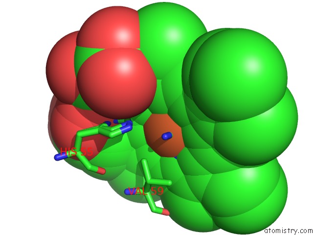
Mono view
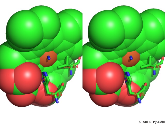
Stereo pair view
Mono view
Stereo pair view
A full contact list of Iron with other atoms in the Fe binding
site number 1 of X-Ray Structure of the Metcyano Form of Dehaloperoxidase From Amphitrite Ornata: Evidence For Photoreductive Lysis of Iron-Cyanide Bond within 5.0Å range:
|
Iron binding site 2 out of 2 in 3kun
Go back to
Iron binding site 2 out
of 2 in the X-Ray Structure of the Metcyano Form of Dehaloperoxidase From Amphitrite Ornata: Evidence For Photoreductive Lysis of Iron-Cyanide Bond
Mono view
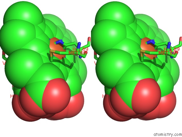
Stereo pair view
Mono view
Stereo pair view
A full contact list of Iron with other atoms in the Fe binding
site number 2 of X-Ray Structure of the Metcyano Form of Dehaloperoxidase From Amphitrite Ornata: Evidence For Photoreductive Lysis of Iron-Cyanide Bond within 5.0Å range:
|
Reference:
V.S.De Serrano,
M.F.Davis,
J.F.Gaff,
Q.Zhang,
Z.Chen,
E.L.D'antonio,
E.F.Bowden,
R.Rose,
S.Franzen.
X-Ray Structure of the Metcyano Form of Dehaloperoxidase From Amphitrite Ornata: Evidence For Photoreductive Dissociation of the Iron-Cyanide Bond. Acta Crystallogr.,Sect.D V. 66 770 2010.
ISSN: ISSN 0907-4449
PubMed: 20606257
DOI: 10.1107/S0907444910014605
Page generated: Sun Aug 4 14:01:06 2024
ISSN: ISSN 0907-4449
PubMed: 20606257
DOI: 10.1107/S0907444910014605
Last articles
Zn in 9MJ5Zn in 9HNW
Zn in 9G0L
Zn in 9FNE
Zn in 9DZN
Zn in 9E0I
Zn in 9D32
Zn in 9DAK
Zn in 8ZXC
Zn in 8ZUF