Iron »
PDB 3kyx-3lhb »
3l4p »
Iron in PDB 3l4p: Crystal Structure of the Aldehyde Dehydrogenase (A.K.A. Aor or Mop) of Desulfovibrio Gigas Covalently Bound to [ASO3]-
Enzymatic activity of Crystal Structure of the Aldehyde Dehydrogenase (A.K.A. Aor or Mop) of Desulfovibrio Gigas Covalently Bound to [ASO3]-
All present enzymatic activity of Crystal Structure of the Aldehyde Dehydrogenase (A.K.A. Aor or Mop) of Desulfovibrio Gigas Covalently Bound to [ASO3]-:
1.2.99.7;
1.2.99.7;
Protein crystallography data
The structure of Crystal Structure of the Aldehyde Dehydrogenase (A.K.A. Aor or Mop) of Desulfovibrio Gigas Covalently Bound to [ASO3]-, PDB code: 3l4p
was solved by
D.R.Boer,
M.J.Romao,
with X-Ray Crystallography technique. A brief refinement statistics is given in the table below:
| Resolution Low / High (Å) | 19.44 / 1.45 |
| Space group | P 61 2 2 |
| Cell size a, b, c (Å), α, β, γ (°) | 142.890, 142.890, 161.640, 90.00, 90.00, 120.00 |
| R / Rfree (%) | 14.2 / 16.6 |
Other elements in 3l4p:
The structure of Crystal Structure of the Aldehyde Dehydrogenase (A.K.A. Aor or Mop) of Desulfovibrio Gigas Covalently Bound to [ASO3]- also contains other interesting chemical elements:
| Molybdenum | (Mo) | 1 atom |
| Magnesium | (Mg) | 5 atoms |
| Arsenic | (As) | 1 atom |
| Calcium | (Ca) | 1 atom |
| Chlorine | (Cl) | 9 atoms |
Iron Binding Sites:
The binding sites of Iron atom in the Crystal Structure of the Aldehyde Dehydrogenase (A.K.A. Aor or Mop) of Desulfovibrio Gigas Covalently Bound to [ASO3]-
(pdb code 3l4p). This binding sites where shown within
5.0 Angstroms radius around Iron atom.
In total 4 binding sites of Iron where determined in the Crystal Structure of the Aldehyde Dehydrogenase (A.K.A. Aor or Mop) of Desulfovibrio Gigas Covalently Bound to [ASO3]-, PDB code: 3l4p:
Jump to Iron binding site number: 1; 2; 3; 4;
In total 4 binding sites of Iron where determined in the Crystal Structure of the Aldehyde Dehydrogenase (A.K.A. Aor or Mop) of Desulfovibrio Gigas Covalently Bound to [ASO3]-, PDB code: 3l4p:
Jump to Iron binding site number: 1; 2; 3; 4;
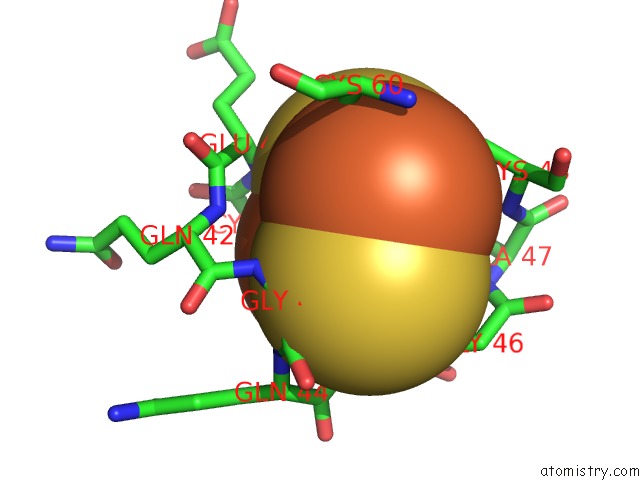
Iron binding site 1 out of 4 in 3l4p
Go back to
Iron binding site 1 out
of 4 in the Crystal Structure of the Aldehyde Dehydrogenase (A.K.A. Aor or Mop) of Desulfovibrio Gigas Covalently Bound to [ASO3]-
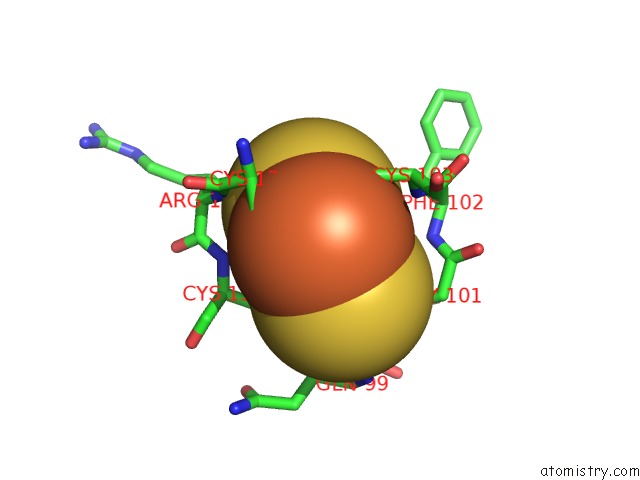
Mono view
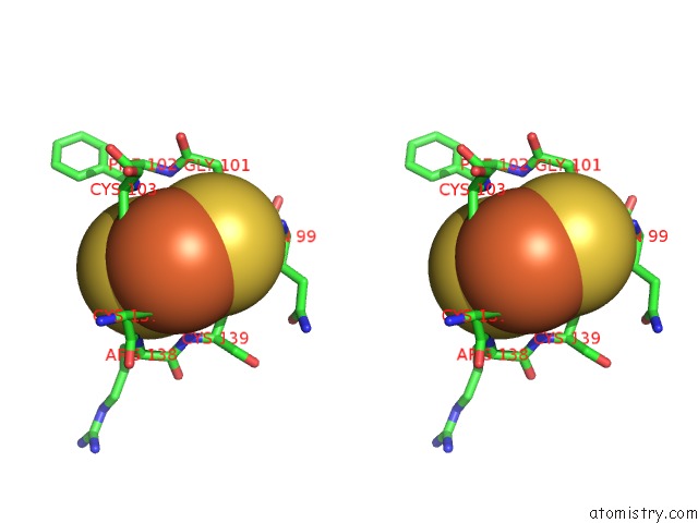
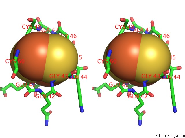
Stereo pair view
Mono view
Stereo pair view
A full contact list of Iron with other atoms in the Fe binding
site number 1 of Crystal Structure of the Aldehyde Dehydrogenase (A.K.A. Aor or Mop) of Desulfovibrio Gigas Covalently Bound to [ASO3]- within 5.0Å range:
|
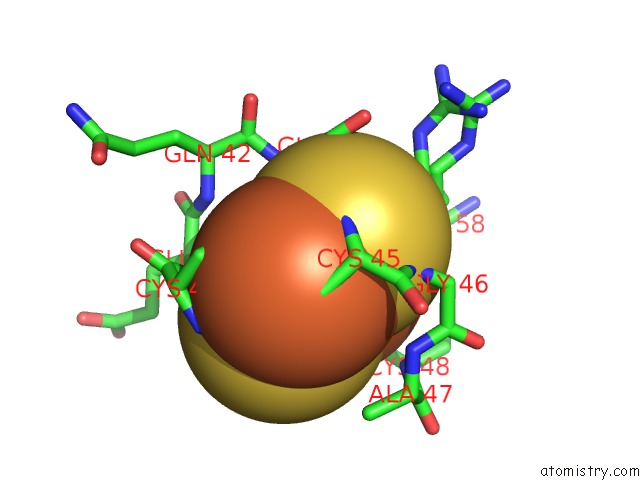
Iron binding site 2 out of 4 in 3l4p
Go back to
Iron binding site 2 out
of 4 in the Crystal Structure of the Aldehyde Dehydrogenase (A.K.A. Aor or Mop) of Desulfovibrio Gigas Covalently Bound to [ASO3]-
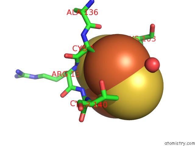
Mono view
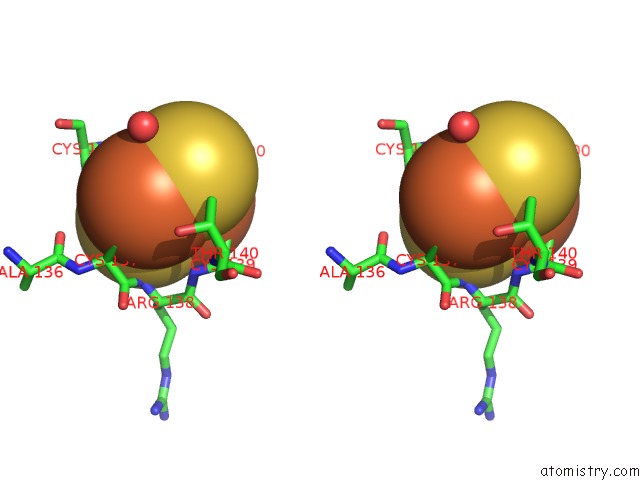
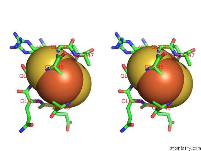
Stereo pair view
Mono view
Stereo pair view
A full contact list of Iron with other atoms in the Fe binding
site number 2 of Crystal Structure of the Aldehyde Dehydrogenase (A.K.A. Aor or Mop) of Desulfovibrio Gigas Covalently Bound to [ASO3]- within 5.0Å range:
|
Iron binding site 3 out of 4 in 3l4p
Go back to
Iron binding site 3 out
of 4 in the Crystal Structure of the Aldehyde Dehydrogenase (A.K.A. Aor or Mop) of Desulfovibrio Gigas Covalently Bound to [ASO3]-
Mono view
Stereo pair view
Mono view
Stereo pair view
A full contact list of Iron with other atoms in the Fe binding
site number 3 of Crystal Structure of the Aldehyde Dehydrogenase (A.K.A. Aor or Mop) of Desulfovibrio Gigas Covalently Bound to [ASO3]- within 5.0Å range:
|
Iron binding site 4 out of 4 in 3l4p
Go back to
Iron binding site 4 out
of 4 in the Crystal Structure of the Aldehyde Dehydrogenase (A.K.A. Aor or Mop) of Desulfovibrio Gigas Covalently Bound to [ASO3]-
Mono view
Stereo pair view
Mono view
Stereo pair view
A full contact list of Iron with other atoms in the Fe binding
site number 4 of Crystal Structure of the Aldehyde Dehydrogenase (A.K.A. Aor or Mop) of Desulfovibrio Gigas Covalently Bound to [ASO3]- within 5.0Å range:
|
Reference:
A.Thapper,
D.R.Boer,
C.D.Brondino,
J.J.Moura,
M.J.Romao.
Correlating Epr and X-Ray Structural Analysis of Arsenite-Inhibited Forms of Aldehyde Oxidoreductase. J.Biol.Inorg.Chem. V. 12 353 2007.
ISSN: ISSN 0949-8257
PubMed: 17139522
DOI: 10.1007/S00775-006-0191-9
Page generated: Sun Aug 4 14:05:17 2024
ISSN: ISSN 0949-8257
PubMed: 17139522
DOI: 10.1007/S00775-006-0191-9
Last articles
F in 4P75F in 4P6G
F in 4P5Z
F in 4OZ3
F in 4P45
F in 4P1U
F in 4P44
F in 4P2H
F in 4OWO
F in 4OZ2