Iron in PDB 3l9q: Crystal Structure of Human Polymerase Alpha-Primase P58 Iron-Sulfur Cluster Domain
Protein crystallography data
The structure of Crystal Structure of Human Polymerase Alpha-Primase P58 Iron-Sulfur Cluster Domain, PDB code: 3l9q
was solved by
S.Vaithiyalingam,
B.F.Eichman,
W.J.Chazin,
with X-Ray Crystallography technique. A brief refinement statistics is given in the table below:
Resolution Low / High (Å)
|
37.32 /
1.70
|
Space group
|
C 1 2 1
|
Cell size a, b, c (Å), α, β, γ (°)
|
109.441,
53.050,
88.844,
90.00,
115.09,
90.00
|
R / Rfree (%)
|
13 /
16.9
|
Iron Binding Sites:
The binding sites of Iron atom in the Crystal Structure of Human Polymerase Alpha-Primase P58 Iron-Sulfur Cluster Domain
(pdb code 3l9q). This binding sites where shown within
5.0 Angstroms radius around Iron atom.
In total 8 binding sites of Iron where determined in the
Crystal Structure of Human Polymerase Alpha-Primase P58 Iron-Sulfur Cluster Domain, PDB code: 3l9q:
Jump to Iron binding site number:
1;
2;
3;
4;
5;
6;
7;
8;
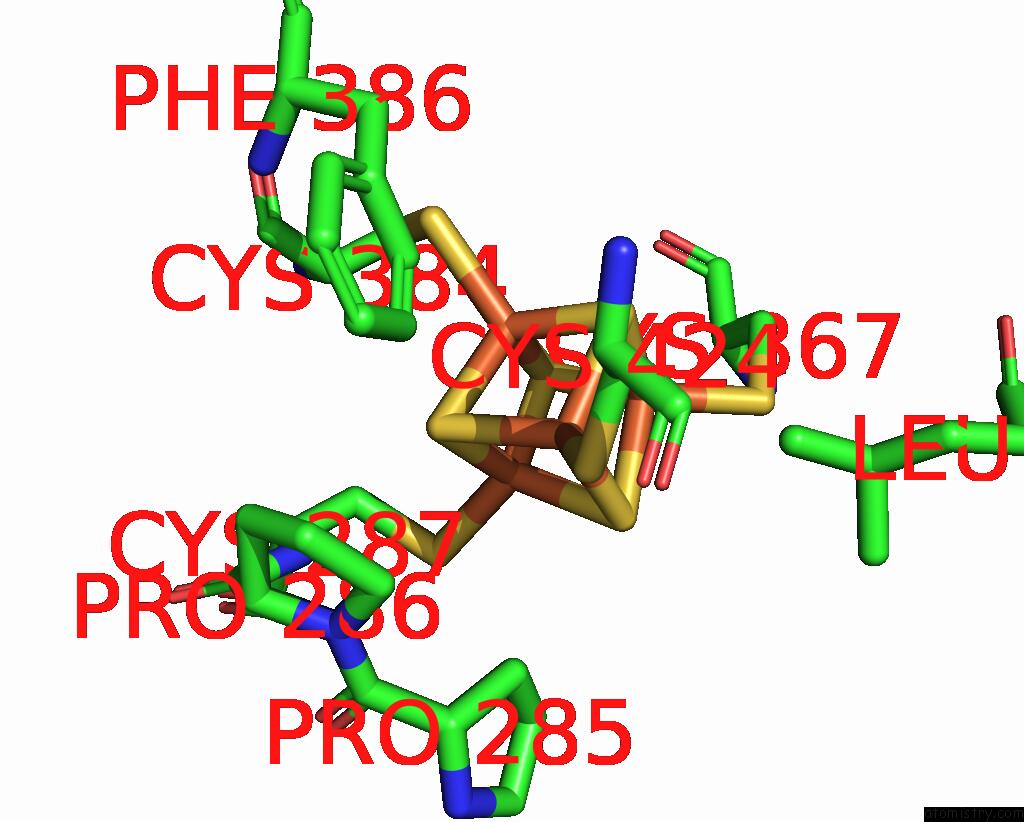
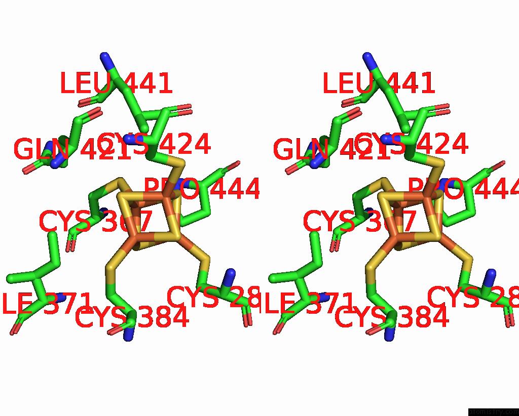
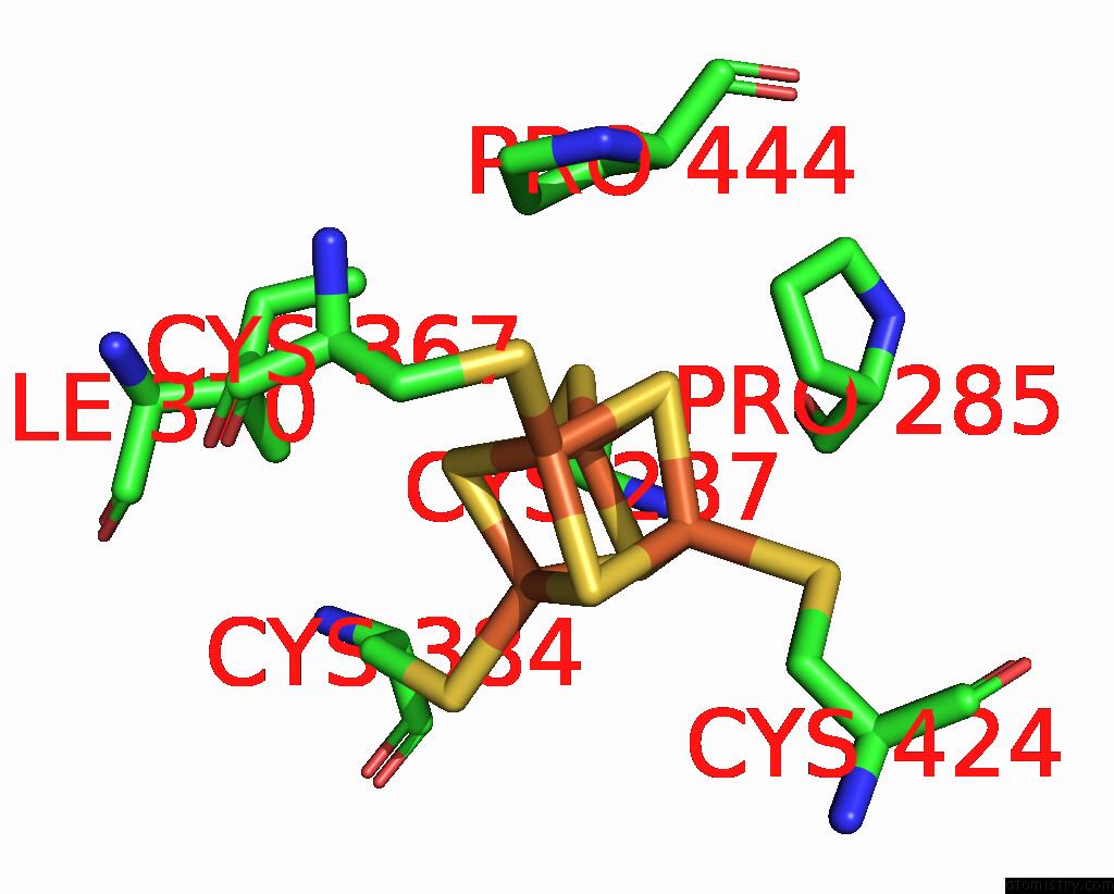
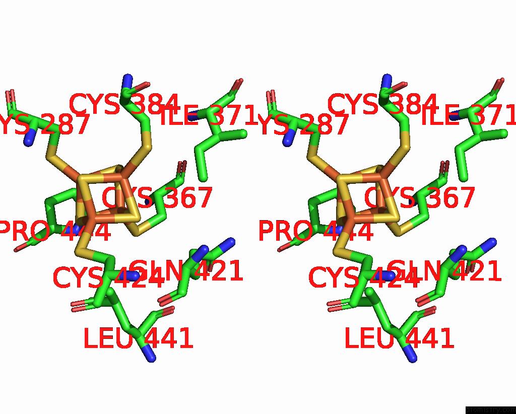
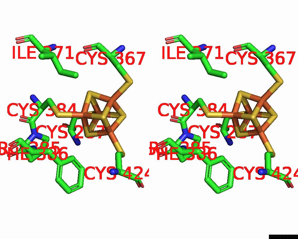
Iron binding site 1 out
of 8 in 3l9q
Go back to
Iron Binding Sites List in 3l9q
Iron binding site 1 out
of 8 in the Crystal Structure of Human Polymerase Alpha-Primase P58 Iron-Sulfur Cluster Domain
 Mono view
 Stereo pair view
|
A full contact list of Iron with other atoms in the Fe binding
site number 1 of Crystal Structure of Human Polymerase Alpha-Primase P58 Iron-Sulfur Cluster Domain within 5.0Å range:
probe
|
atom
|
residue
|
distance (Å)
|
B
|
Occ
|
A:Fe500
b:22.0
occ:1.00
|
FE1
|
A:SF4500
|
0.0
|
22.0
|
1.0
|
S3
|
A:SF4500
|
2.3
|
21.4
|
1.0
|
S2
|
A:SF4500
|
2.3
|
21.2
|
1.0
|
S4
|
A:SF4500
|
2.3
|
21.4
|
1.0
|
SG
|
A:CYS424
|
2.3
|
22.0
|
1.0
|
FE4
|
A:SF4500
|
2.7
|
20.7
|
1.0
|
FE2
|
A:SF4500
|
2.7
|
21.7
|
1.0
|
FE3
|
A:SF4500
|
2.7
|
20.7
|
1.0
|
CB
|
A:CYS424
|
3.1
|
20.9
|
1.0
|
S1
|
A:SF4500
|
3.9
|
21.0
|
1.0
|
CD2
|
A:LEU441
|
3.9
|
23.7
|
1.0
|
CB
|
A:PRO285
|
4.2
|
18.1
|
1.0
|
CE2
|
A:PHE386
|
4.3
|
24.8
|
1.0
|
CD
|
A:PRO286
|
4.4
|
17.4
|
1.0
|
CD2
|
A:PHE386
|
4.4
|
24.4
|
1.0
|
CA
|
A:CYS424
|
4.6
|
21.9
|
1.0
|
SG
|
A:CYS367
|
4.7
|
22.7
|
1.0
|
SG
|
A:CYS287
|
4.7
|
19.3
|
1.0
|
SG
|
A:CYS384
|
4.8
|
21.7
|
1.0
|
|
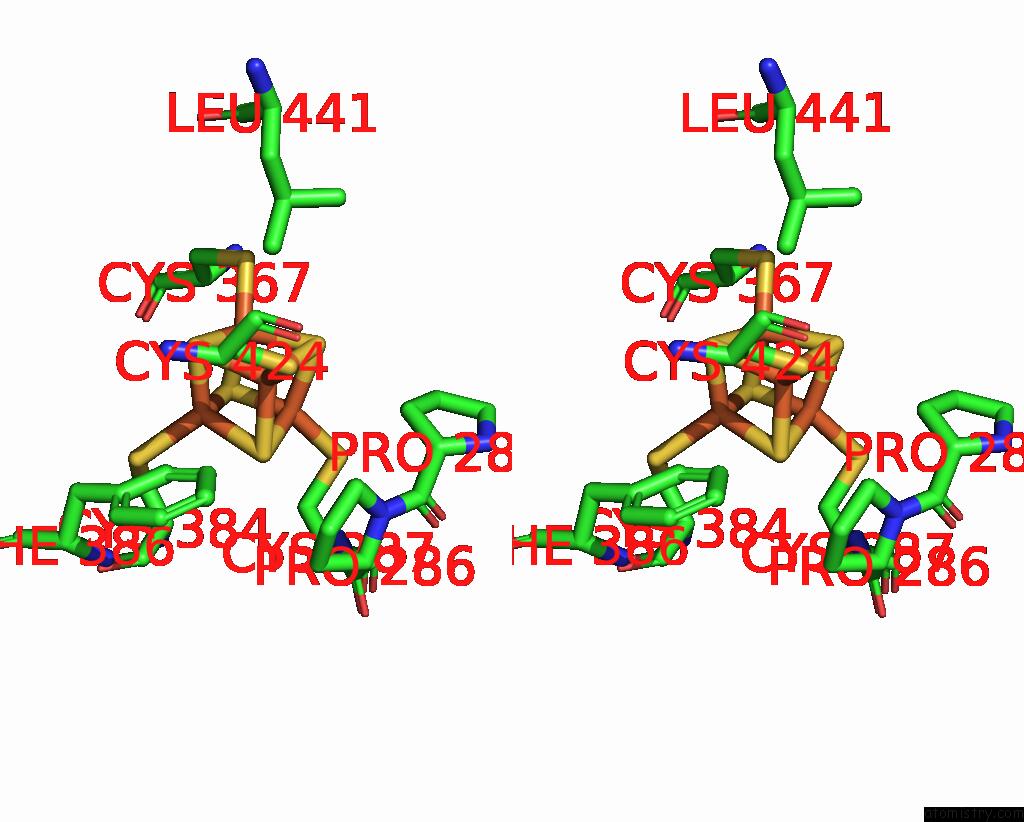
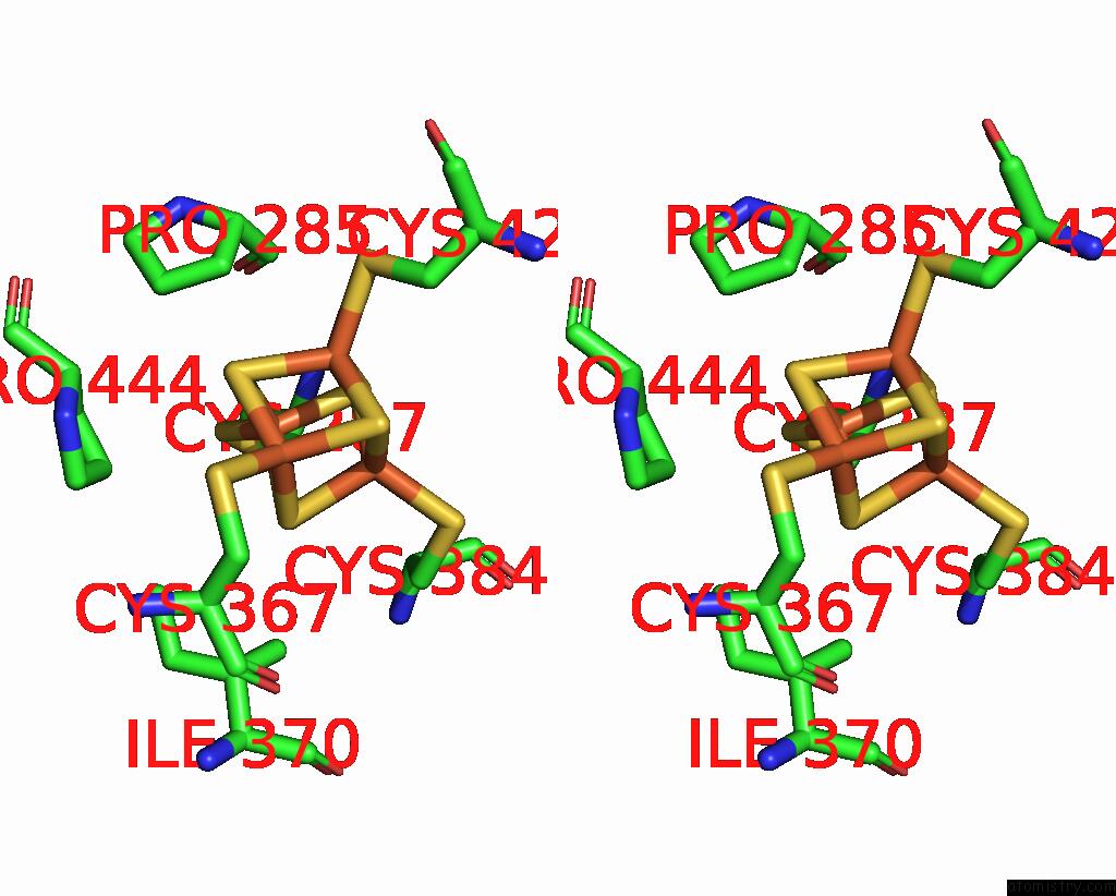
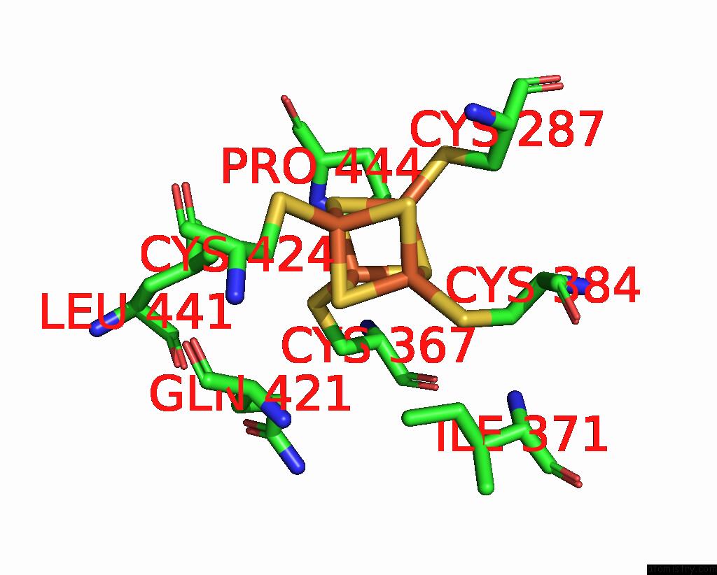
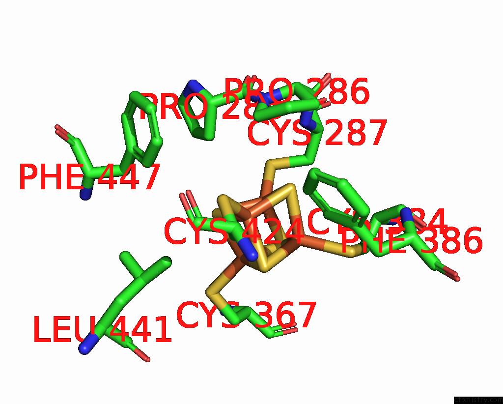
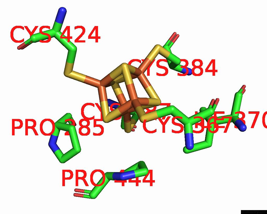
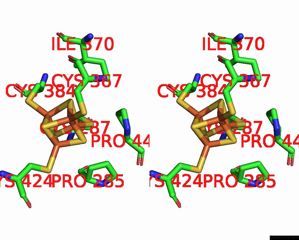
Iron binding site 2 out
of 8 in 3l9q
Go back to
Iron Binding Sites List in 3l9q
Iron binding site 2 out
of 8 in the Crystal Structure of Human Polymerase Alpha-Primase P58 Iron-Sulfur Cluster Domain
 Mono view
 Stereo pair view
|
A full contact list of Iron with other atoms in the Fe binding
site number 2 of Crystal Structure of Human Polymerase Alpha-Primase P58 Iron-Sulfur Cluster Domain within 5.0Å range:
probe
|
atom
|
residue
|
distance (Å)
|
B
|
Occ
|
A:Fe500
b:21.7
occ:1.00
|
FE2
|
A:SF4500
|
0.0
|
21.7
|
1.0
|
S4
|
A:SF4500
|
2.3
|
21.4
|
1.0
|
S1
|
A:SF4500
|
2.3
|
21.0
|
1.0
|
S3
|
A:SF4500
|
2.3
|
21.4
|
1.0
|
SG
|
A:CYS384
|
2.3
|
21.7
|
1.0
|
FE4
|
A:SF4500
|
2.7
|
20.7
|
1.0
|
FE1
|
A:SF4500
|
2.7
|
22.0
|
1.0
|
FE3
|
A:SF4500
|
2.7
|
20.7
|
1.0
|
CB
|
A:CYS384
|
3.3
|
21.1
|
1.0
|
S2
|
A:SF4500
|
3.9
|
21.2
|
1.0
|
CA
|
A:CYS384
|
4.1
|
19.6
|
1.0
|
CD2
|
A:PHE386
|
4.2
|
24.4
|
1.0
|
CD1
|
A:ILE371
|
4.6
|
24.2
|
1.0
|
CB
|
A:CYS287
|
4.6
|
19.0
|
1.0
|
CD
|
A:PRO385
|
4.7
|
22.4
|
1.0
|
SG
|
A:CYS287
|
4.7
|
19.3
|
1.0
|
CG1
|
A:ILE371
|
4.7
|
22.8
|
1.0
|
CE2
|
A:PHE386
|
4.8
|
24.8
|
1.0
|
C
|
A:CYS384
|
4.8
|
20.4
|
1.0
|
SG
|
A:CYS424
|
4.8
|
22.0
|
1.0
|
CB
|
A:CYS424
|
4.9
|
20.9
|
1.0
|
SG
|
A:CYS367
|
5.0
|
22.7
|
1.0
|
N
|
A:PRO385
|
5.0
|
20.6
|
1.0
|
|
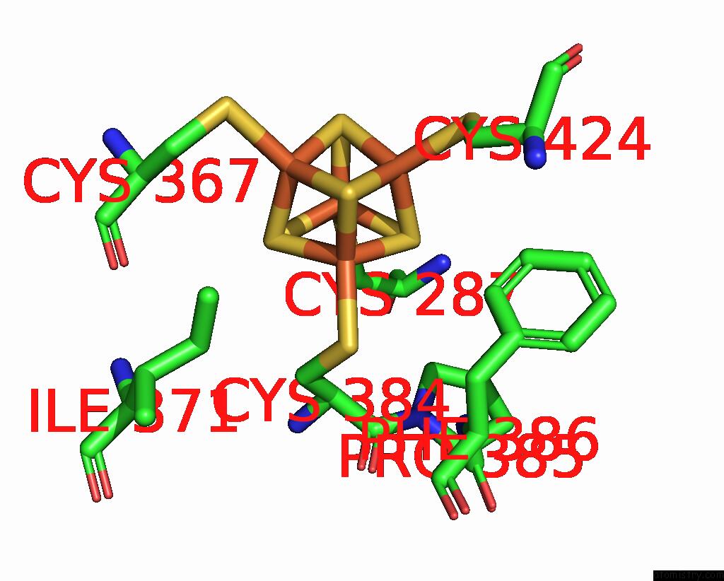
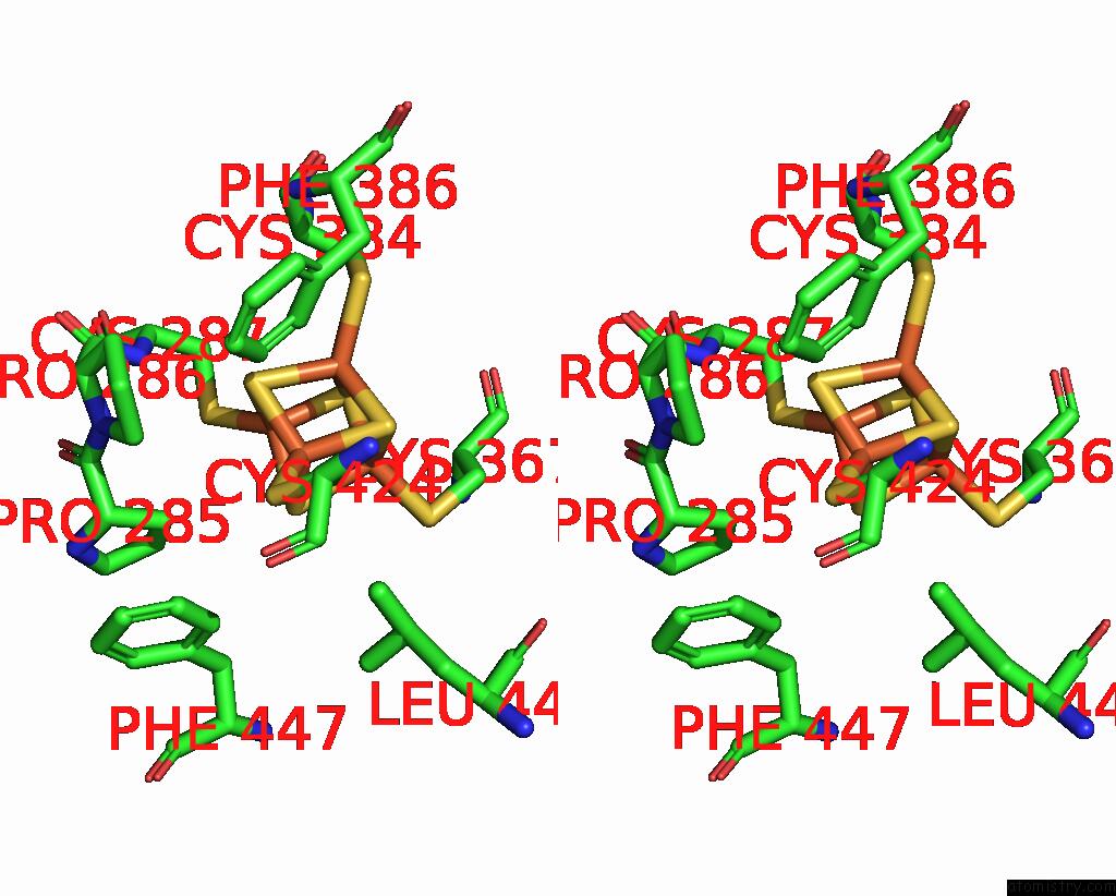
Iron binding site 3 out
of 8 in 3l9q
Go back to
Iron Binding Sites List in 3l9q
Iron binding site 3 out
of 8 in the Crystal Structure of Human Polymerase Alpha-Primase P58 Iron-Sulfur Cluster Domain
 Mono view
 Stereo pair view
|
A full contact list of Iron with other atoms in the Fe binding
site number 3 of Crystal Structure of Human Polymerase Alpha-Primase P58 Iron-Sulfur Cluster Domain within 5.0Å range:
probe
|
atom
|
residue
|
distance (Å)
|
B
|
Occ
|
A:Fe500
b:20.7
occ:1.00
|
FE3
|
A:SF4500
|
0.0
|
20.7
|
1.0
|
S1
|
A:SF4500
|
2.3
|
21.0
|
1.0
|
S2
|
A:SF4500
|
2.3
|
21.2
|
1.0
|
S4
|
A:SF4500
|
2.3
|
21.4
|
1.0
|
SG
|
A:CYS367
|
2.3
|
22.7
|
1.0
|
FE4
|
A:SF4500
|
2.7
|
20.7
|
1.0
|
FE1
|
A:SF4500
|
2.7
|
22.0
|
1.0
|
FE2
|
A:SF4500
|
2.7
|
21.7
|
1.0
|
CB
|
A:CYS367
|
3.2
|
21.6
|
1.0
|
CA
|
A:CYS367
|
3.8
|
21.3
|
1.0
|
S3
|
A:SF4500
|
3.9
|
21.4
|
1.0
|
CD2
|
A:LEU441
|
4.3
|
23.7
|
1.0
|
CG
|
A:PRO444
|
4.5
|
15.4
|
1.0
|
CD
|
A:PRO444
|
4.6
|
15.3
|
1.0
|
CG
|
A:GLN421
|
4.6
|
25.8
|
1.0
|
N
|
A:CYS367
|
4.8
|
19.5
|
1.0
|
SG
|
A:CYS424
|
4.8
|
22.0
|
1.0
|
CG
|
A:LEU441
|
4.8
|
22.1
|
1.0
|
N
|
A:PRO444
|
4.8
|
16.4
|
1.0
|
SG
|
A:CYS287
|
4.9
|
19.3
|
1.0
|
SG
|
A:CYS384
|
4.9
|
21.7
|
1.0
|
CD1
|
A:ILE371
|
4.9
|
24.2
|
1.0
|
C
|
A:CYS367
|
4.9
|
22.3
|
1.0
|
|
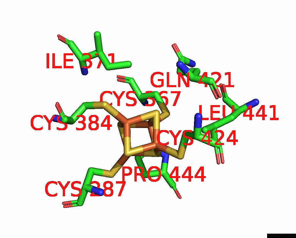
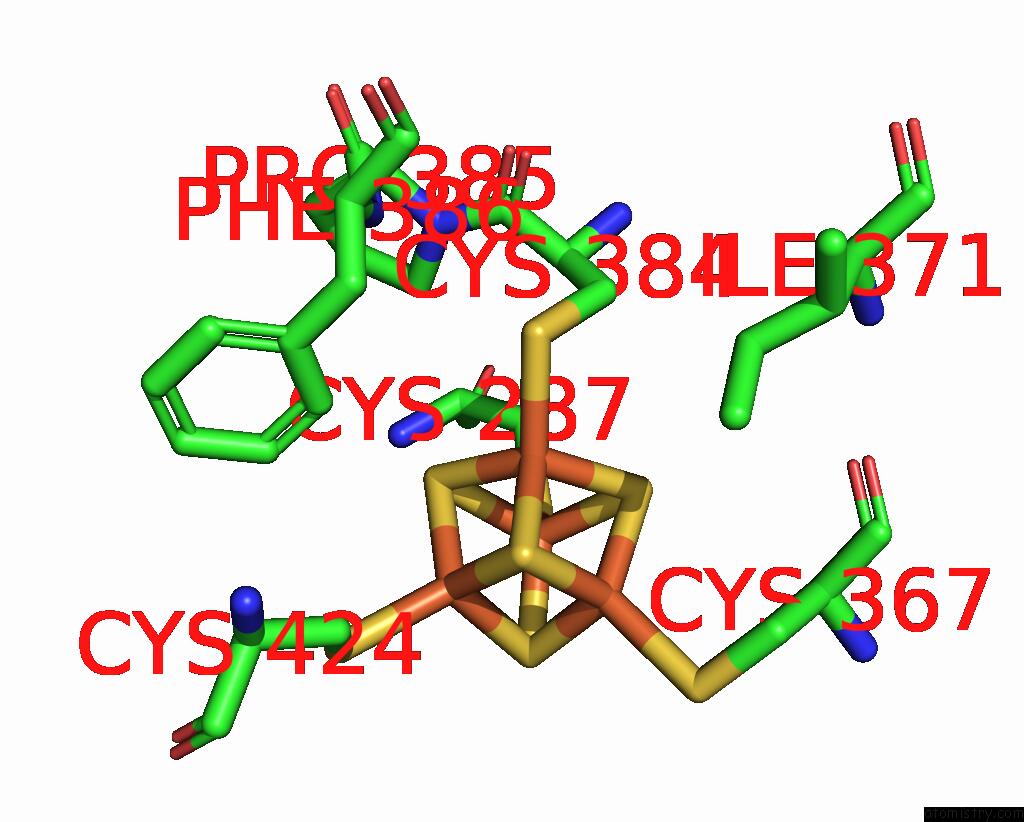
Iron binding site 4 out
of 8 in 3l9q
Go back to
Iron Binding Sites List in 3l9q
Iron binding site 4 out
of 8 in the Crystal Structure of Human Polymerase Alpha-Primase P58 Iron-Sulfur Cluster Domain
 Mono view
 Stereo pair view
|
A full contact list of Iron with other atoms in the Fe binding
site number 4 of Crystal Structure of Human Polymerase Alpha-Primase P58 Iron-Sulfur Cluster Domain within 5.0Å range:
probe
|
atom
|
residue
|
distance (Å)
|
B
|
Occ
|
A:Fe500
b:20.7
occ:1.00
|
FE4
|
A:SF4500
|
0.0
|
20.7
|
1.0
|
S2
|
A:SF4500
|
2.3
|
21.2
|
1.0
|
SG
|
A:CYS287
|
2.3
|
19.3
|
1.0
|
S3
|
A:SF4500
|
2.3
|
21.4
|
1.0
|
S1
|
A:SF4500
|
2.3
|
21.0
|
1.0
|
FE2
|
A:SF4500
|
2.7
|
21.7
|
1.0
|
FE1
|
A:SF4500
|
2.7
|
22.0
|
1.0
|
FE3
|
A:SF4500
|
2.7
|
20.7
|
1.0
|
CB
|
A:CYS287
|
3.2
|
19.0
|
1.0
|
S4
|
A:SF4500
|
3.9
|
21.4
|
1.0
|
CB
|
A:PRO285
|
4.1
|
18.1
|
1.0
|
N
|
A:CYS287
|
4.1
|
17.2
|
1.0
|
CG
|
A:PRO444
|
4.1
|
15.4
|
1.0
|
CA
|
A:CYS287
|
4.3
|
19.0
|
1.0
|
CG
|
A:PRO285
|
4.4
|
17.3
|
1.0
|
CB
|
A:PRO444
|
4.7
|
14.9
|
1.0
|
SG
|
A:CYS367
|
4.7
|
22.7
|
1.0
|
SG
|
A:CYS384
|
4.8
|
21.7
|
1.0
|
CD1
|
A:ILE370
|
4.8
|
20.4
|
1.0
|
SG
|
A:CYS424
|
4.9
|
22.0
|
1.0
|
|
Iron binding site 5 out
of 8 in 3l9q
Go back to
Iron Binding Sites List in 3l9q
Iron binding site 5 out
of 8 in the Crystal Structure of Human Polymerase Alpha-Primase P58 Iron-Sulfur Cluster Domain
 Mono view
 Stereo pair view
|
A full contact list of Iron with other atoms in the Fe binding
site number 5 of Crystal Structure of Human Polymerase Alpha-Primase P58 Iron-Sulfur Cluster Domain within 5.0Å range:
probe
|
atom
|
residue
|
distance (Å)
|
B
|
Occ
|
B:Fe500
b:16.1
occ:1.00
|
FE1
|
B:SF4500
|
0.0
|
16.1
|
1.0
|
S2
|
B:SF4500
|
2.3
|
15.6
|
1.0
|
S4
|
B:SF4500
|
2.3
|
16.0
|
1.0
|
S3
|
B:SF4500
|
2.3
|
16.2
|
1.0
|
SG
|
B:CYS367
|
2.3
|
15.7
|
1.0
|
FE4
|
B:SF4500
|
2.7
|
15.6
|
1.0
|
FE2
|
B:SF4500
|
2.7
|
15.1
|
1.0
|
FE3
|
B:SF4500
|
2.8
|
15.4
|
1.0
|
CB
|
B:CYS367
|
3.2
|
16.6
|
1.0
|
CA
|
B:CYS367
|
3.8
|
16.1
|
1.0
|
S1
|
B:SF4500
|
3.9
|
15.1
|
1.0
|
CG
|
B:PRO444
|
4.4
|
13.9
|
1.0
|
CD
|
B:PRO444
|
4.5
|
15.4
|
1.0
|
CD2
|
B:LEU441
|
4.6
|
20.2
|
1.0
|
CG
|
B:GLN421
|
4.7
|
18.6
|
1.0
|
N
|
B:CYS367
|
4.7
|
15.1
|
1.0
|
SG
|
B:CYS424
|
4.8
|
15.5
|
1.0
|
N
|
B:PRO444
|
4.8
|
14.8
|
1.0
|
SG
|
B:CYS384
|
4.9
|
16.0
|
1.0
|
SG
|
B:CYS287
|
4.9
|
16.0
|
1.0
|
CD1
|
B:ILE371
|
4.9
|
16.3
|
1.0
|
C
|
B:CYS367
|
4.9
|
18.2
|
1.0
|
|
Iron binding site 6 out
of 8 in 3l9q
Go back to
Iron Binding Sites List in 3l9q
Iron binding site 6 out
of 8 in the Crystal Structure of Human Polymerase Alpha-Primase P58 Iron-Sulfur Cluster Domain
 Mono view
 Stereo pair view
|
A full contact list of Iron with other atoms in the Fe binding
site number 6 of Crystal Structure of Human Polymerase Alpha-Primase P58 Iron-Sulfur Cluster Domain within 5.0Å range:
probe
|
atom
|
residue
|
distance (Å)
|
B
|
Occ
|
B:Fe500
b:15.1
occ:1.00
|
FE2
|
B:SF4500
|
0.0
|
15.1
|
1.0
|
S1
|
B:SF4500
|
2.3
|
15.1
|
1.0
|
S3
|
B:SF4500
|
2.3
|
16.2
|
1.0
|
S4
|
B:SF4500
|
2.3
|
16.0
|
1.0
|
SG
|
B:CYS424
|
2.3
|
15.5
|
1.0
|
FE4
|
B:SF4500
|
2.7
|
15.6
|
1.0
|
FE3
|
B:SF4500
|
2.7
|
15.4
|
1.0
|
FE1
|
B:SF4500
|
2.7
|
16.1
|
1.0
|
CB
|
B:CYS424
|
3.2
|
15.1
|
1.0
|
S2
|
B:SF4500
|
3.9
|
15.6
|
1.0
|
CD2
|
B:LEU441
|
4.2
|
20.2
|
1.0
|
CB
|
B:PRO285
|
4.2
|
15.5
|
1.0
|
CE2
|
B:PHE386
|
4.2
|
15.1
|
1.0
|
CD2
|
B:PHE386
|
4.4
|
15.8
|
1.0
|
CD
|
B:PRO286
|
4.5
|
14.6
|
1.0
|
CA
|
B:CYS424
|
4.6
|
15.5
|
1.0
|
SG
|
B:CYS367
|
4.8
|
15.7
|
1.0
|
SG
|
B:CYS287
|
4.8
|
16.0
|
1.0
|
SG
|
B:CYS384
|
4.8
|
16.0
|
1.0
|
CD1
|
B:PHE447
|
5.0
|
16.2
|
1.0
|
|
Iron binding site 7 out
of 8 in 3l9q
Go back to
Iron Binding Sites List in 3l9q
Iron binding site 7 out
of 8 in the Crystal Structure of Human Polymerase Alpha-Primase P58 Iron-Sulfur Cluster Domain
 Mono view
 Stereo pair view
|
A full contact list of Iron with other atoms in the Fe binding
site number 7 of Crystal Structure of Human Polymerase Alpha-Primase P58 Iron-Sulfur Cluster Domain within 5.0Å range:
probe
|
atom
|
residue
|
distance (Å)
|
B
|
Occ
|
B:Fe500
b:15.4
occ:1.00
|
FE3
|
B:SF4500
|
0.0
|
15.4
|
1.0
|
S4
|
B:SF4500
|
2.3
|
16.0
|
1.0
|
S2
|
B:SF4500
|
2.3
|
15.6
|
1.0
|
S1
|
B:SF4500
|
2.3
|
15.1
|
1.0
|
SG
|
B:CYS384
|
2.3
|
16.0
|
1.0
|
FE4
|
B:SF4500
|
2.7
|
15.6
|
1.0
|
FE2
|
B:SF4500
|
2.7
|
15.1
|
1.0
|
FE1
|
B:SF4500
|
2.8
|
16.1
|
1.0
|
CB
|
B:CYS384
|
3.3
|
15.6
|
1.0
|
S3
|
B:SF4500
|
3.9
|
16.2
|
1.0
|
CA
|
B:CYS384
|
4.1
|
14.3
|
1.0
|
CD2
|
B:PHE386
|
4.1
|
15.8
|
1.0
|
CB
|
B:CYS287
|
4.6
|
16.4
|
1.0
|
CD1
|
B:ILE371
|
4.6
|
16.3
|
1.0
|
SG
|
B:CYS287
|
4.7
|
16.0
|
1.0
|
CG1
|
B:ILE371
|
4.7
|
15.9
|
1.0
|
CD
|
B:PRO385
|
4.7
|
17.9
|
1.0
|
C
|
B:CYS384
|
4.7
|
15.1
|
1.0
|
CE2
|
B:PHE386
|
4.8
|
15.1
|
1.0
|
SG
|
B:CYS424
|
4.8
|
15.5
|
1.0
|
N
|
B:PRO385
|
4.9
|
14.9
|
1.0
|
CB
|
B:CYS424
|
5.0
|
15.1
|
1.0
|
SG
|
B:CYS367
|
5.0
|
15.7
|
1.0
|
|
Iron binding site 8 out
of 8 in 3l9q
Go back to
Iron Binding Sites List in 3l9q
Iron binding site 8 out
of 8 in the Crystal Structure of Human Polymerase Alpha-Primase P58 Iron-Sulfur Cluster Domain
 Mono view
 Stereo pair view
|
A full contact list of Iron with other atoms in the Fe binding
site number 8 of Crystal Structure of Human Polymerase Alpha-Primase P58 Iron-Sulfur Cluster Domain within 5.0Å range:
probe
|
atom
|
residue
|
distance (Å)
|
B
|
Occ
|
B:Fe500
b:15.6
occ:1.00
|
FE4
|
B:SF4500
|
0.0
|
15.6
|
1.0
|
S3
|
B:SF4500
|
2.3
|
16.2
|
1.0
|
SG
|
B:CYS287
|
2.3
|
16.0
|
1.0
|
S2
|
B:SF4500
|
2.3
|
15.6
|
1.0
|
S1
|
B:SF4500
|
2.3
|
15.1
|
1.0
|
FE3
|
B:SF4500
|
2.7
|
15.4
|
1.0
|
FE2
|
B:SF4500
|
2.7
|
15.1
|
1.0
|
FE1
|
B:SF4500
|
2.7
|
16.1
|
1.0
|
CB
|
B:CYS287
|
3.2
|
16.4
|
1.0
|
S4
|
B:SF4500
|
3.9
|
16.0
|
1.0
|
CG
|
B:PRO444
|
4.0
|
13.9
|
1.0
|
CB
|
B:PRO285
|
4.1
|
15.5
|
1.0
|
N
|
B:CYS287
|
4.1
|
15.2
|
1.0
|
CA
|
B:CYS287
|
4.2
|
17.0
|
1.0
|
CG
|
B:PRO285
|
4.4
|
15.7
|
1.0
|
CB
|
B:PRO444
|
4.5
|
14.3
|
1.0
|
SG
|
B:CYS384
|
4.7
|
16.0
|
1.0
|
SG
|
B:CYS367
|
4.8
|
15.7
|
1.0
|
SG
|
B:CYS424
|
4.9
|
15.5
|
1.0
|
CA
|
B:PRO444
|
4.9
|
14.0
|
1.0
|
CD1
|
B:ILE370
|
5.0
|
15.8
|
1.0
|
CD
|
B:PRO444
|
5.0
|
15.4
|
1.0
|
CB
|
B:CYS384
|
5.0
|
15.6
|
1.0
|
|
Reference:
S.Vaithiyalingam,
E.M.Warren,
B.F.Eichman,
W.J.Chazin.
Insights Into Eukaryotic Dna Priming From the Structure and Functional Interactions of the 4FE-4S Cluster Domain of Human Dna Primase. Proc.Natl.Acad.Sci.Usa V. 107 13684 2010.
ISSN: ISSN 0027-8424
PubMed: 20643958
DOI: 10.1073/PNAS.1002009107
Page generated: Sun Aug 4 14:09:59 2024
|