Iron »
PDB 3kyx-3lhb »
3lah »
Iron in PDB 3lah: Structural Insights Into the Molecular Mechanism of H-Nox Activation
Protein crystallography data
The structure of Structural Insights Into the Molecular Mechanism of H-Nox Activation, PDB code: 3lah
was solved by
C.Olea Jr,
M.A.Herzik Jr,
J.Kuriyan,
M.A.Marletta,
with X-Ray Crystallography technique. A brief refinement statistics is given in the table below:
| Resolution Low / High (Å) | 44.60 / 2.00 |
| Space group | P 21 21 21 |
| Cell size a, b, c (Å), α, β, γ (°) | 61.353, 88.713, 89.207, 90.00, 90.00, 90.00 |
| R / Rfree (%) | 21.5 / 25.1 |
Iron Binding Sites:
The binding sites of Iron atom in the Structural Insights Into the Molecular Mechanism of H-Nox Activation
(pdb code 3lah). This binding sites where shown within
5.0 Angstroms radius around Iron atom.
In total 2 binding sites of Iron where determined in the Structural Insights Into the Molecular Mechanism of H-Nox Activation, PDB code: 3lah:
Jump to Iron binding site number: 1; 2;
In total 2 binding sites of Iron where determined in the Structural Insights Into the Molecular Mechanism of H-Nox Activation, PDB code: 3lah:
Jump to Iron binding site number: 1; 2;
Iron binding site 1 out of 2 in 3lah
Go back to
Iron binding site 1 out
of 2 in the Structural Insights Into the Molecular Mechanism of H-Nox Activation
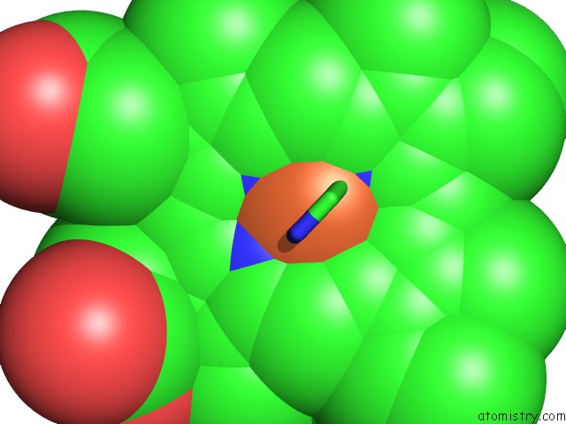
Mono view
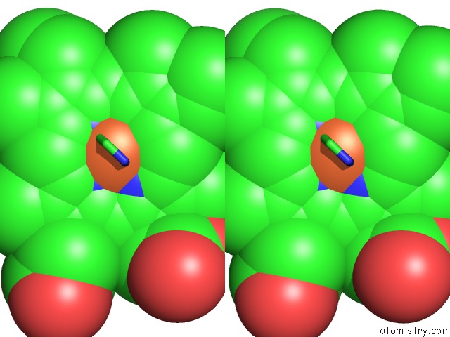
Stereo pair view
Mono view
Stereo pair view
A full contact list of Iron with other atoms in the Fe binding
site number 1 of Structural Insights Into the Molecular Mechanism of H-Nox Activation within 5.0Å range:
|
Iron binding site 2 out of 2 in 3lah
Go back to
Iron binding site 2 out
of 2 in the Structural Insights Into the Molecular Mechanism of H-Nox Activation
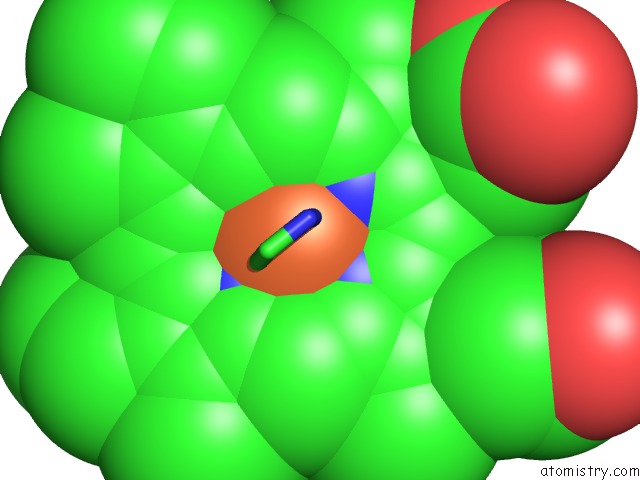
Mono view
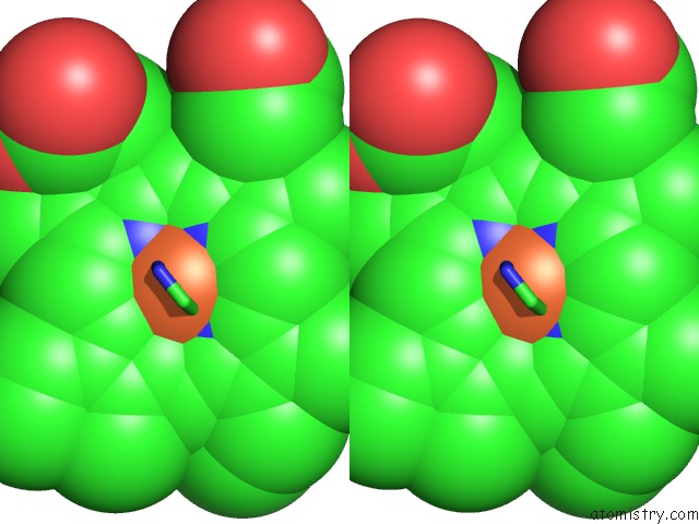
Stereo pair view
Mono view
Stereo pair view
A full contact list of Iron with other atoms in the Fe binding
site number 2 of Structural Insights Into the Molecular Mechanism of H-Nox Activation within 5.0Å range:
|
Reference:
C.Olea,
M.A.Herzik,
J.Kuriyan,
M.A.Marletta.
Structural Insights Into the Molecular Mechanism of H-Nox Activation. Protein Sci. V. 19 881 2010.
ISSN: ISSN 0961-8368
PubMed: 20162612
DOI: 10.1002/PRO.357
Page generated: Sun Aug 4 14:10:02 2024
ISSN: ISSN 0961-8368
PubMed: 20162612
DOI: 10.1002/PRO.357
Last articles
Zn in 9J0NZn in 9J0O
Zn in 9J0P
Zn in 9FJX
Zn in 9EKB
Zn in 9C0F
Zn in 9CAH
Zn in 9CH0
Zn in 9CH3
Zn in 9CH1