Iron »
PDB 3kyx-3lhb »
3lb8 »
Iron in PDB 3lb8: Crystal Structure of the Covalent Putidaredoxin Reductase- Putidaredoxin Complex
Protein crystallography data
The structure of Crystal Structure of the Covalent Putidaredoxin Reductase- Putidaredoxin Complex, PDB code: 3lb8
was solved by
I.F.Sevrioukova,
with X-Ray Crystallography technique. A brief refinement statistics is given in the table below:
| Resolution Low / High (Å) | 49.50 / 2.60 |
| Space group | P 21 21 21 |
| Cell size a, b, c (Å), α, β, γ (°) | 67.600, 103.400, 167.700, 90.00, 90.00, 90.00 |
| R / Rfree (%) | 24.4 / 27.2 |
Iron Binding Sites:
The binding sites of Iron atom in the Crystal Structure of the Covalent Putidaredoxin Reductase- Putidaredoxin Complex
(pdb code 3lb8). This binding sites where shown within
5.0 Angstroms radius around Iron atom.
In total 4 binding sites of Iron where determined in the Crystal Structure of the Covalent Putidaredoxin Reductase- Putidaredoxin Complex, PDB code: 3lb8:
Jump to Iron binding site number: 1; 2; 3; 4;
In total 4 binding sites of Iron where determined in the Crystal Structure of the Covalent Putidaredoxin Reductase- Putidaredoxin Complex, PDB code: 3lb8:
Jump to Iron binding site number: 1; 2; 3; 4;
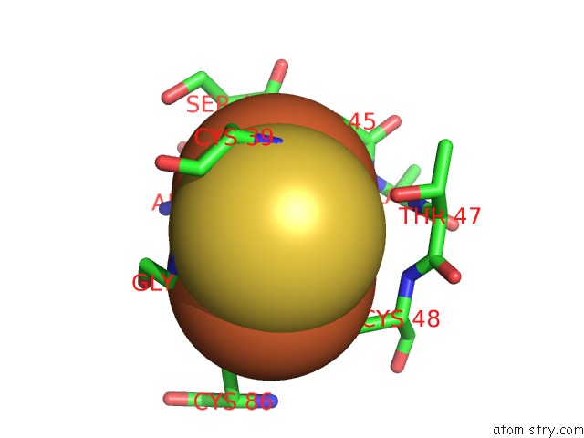
Iron binding site 1 out of 4 in 3lb8
Go back to
Iron binding site 1 out
of 4 in the Crystal Structure of the Covalent Putidaredoxin Reductase- Putidaredoxin Complex
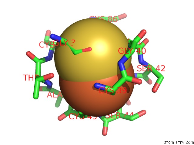
Mono view
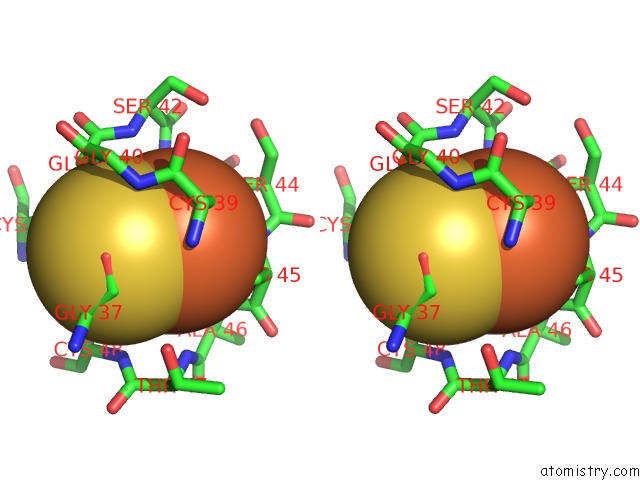
Stereo pair view
Mono view
Stereo pair view
A full contact list of Iron with other atoms in the Fe binding
site number 1 of Crystal Structure of the Covalent Putidaredoxin Reductase- Putidaredoxin Complex within 5.0Å range:
|
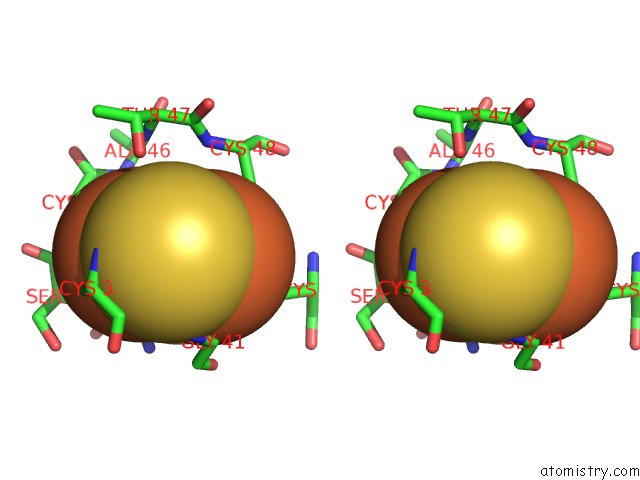
Iron binding site 2 out of 4 in 3lb8
Go back to
Iron binding site 2 out
of 4 in the Crystal Structure of the Covalent Putidaredoxin Reductase- Putidaredoxin Complex
Mono view
Stereo pair view
Mono view
Stereo pair view
A full contact list of Iron with other atoms in the Fe binding
site number 2 of Crystal Structure of the Covalent Putidaredoxin Reductase- Putidaredoxin Complex within 5.0Å range:
|
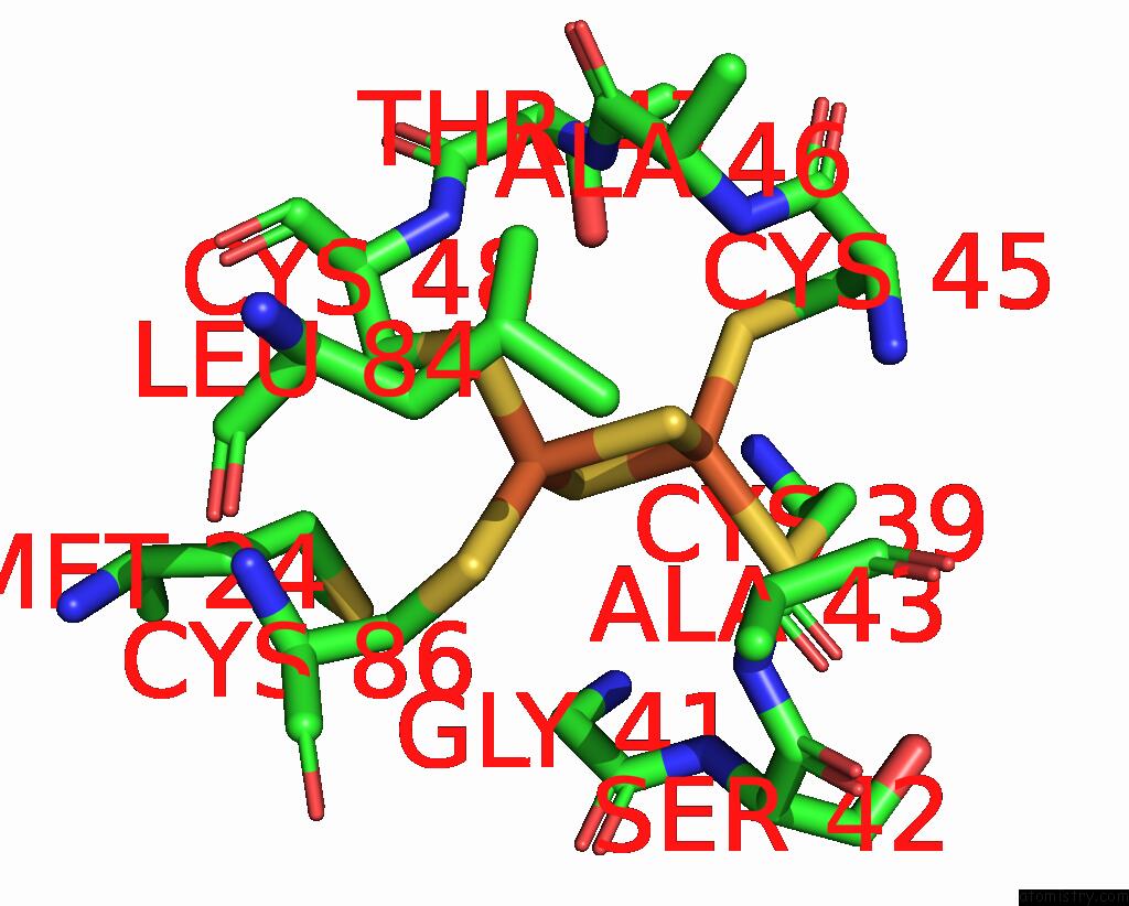
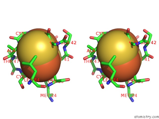
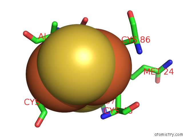
Iron binding site 3 out of 4 in 3lb8
Go back to
Iron binding site 3 out
of 4 in the Crystal Structure of the Covalent Putidaredoxin Reductase- Putidaredoxin Complex
Mono view
Stereo pair view
Mono view
Stereo pair view
A full contact list of Iron with other atoms in the Fe binding
site number 3 of Crystal Structure of the Covalent Putidaredoxin Reductase- Putidaredoxin Complex within 5.0Å range:
|
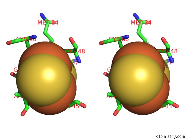
Iron binding site 4 out of 4 in 3lb8
Go back to
Iron binding site 4 out
of 4 in the Crystal Structure of the Covalent Putidaredoxin Reductase- Putidaredoxin Complex
Mono view
Stereo pair view
Mono view
Stereo pair view
A full contact list of Iron with other atoms in the Fe binding
site number 4 of Crystal Structure of the Covalent Putidaredoxin Reductase- Putidaredoxin Complex within 5.0Å range:
|
Reference:
I.F.Sevrioukova,
T.L.Poulos,
I.Y.Churbanova.
Crystal Structure of the Putidaredoxin Reductase X Putidaredoxin Electron Transfer Complex. J.Biol.Chem. V. 285 13616 2010.
ISSN: ISSN 0021-9258
PubMed: 20179327
DOI: 10.1074/JBC.M110.104968
Page generated: Sun Aug 4 14:16:40 2024
ISSN: ISSN 0021-9258
PubMed: 20179327
DOI: 10.1074/JBC.M110.104968
Last articles
Cl in 5XTUCl in 5XNP
Cl in 5XPF
Cl in 5XS2
Cl in 5XRB
Cl in 5XR5
Cl in 5XQE
Cl in 5XQD
Cl in 5XPE
Cl in 5XNL