Iron »
PDB 3kyx-3lhb »
3lhb »
Iron in PDB 3lhb: The 2.7 Angstrom Crystal Structure of Deoxygenated Hemoglobin From the Sea Lamprey (Petromyzon Marinus)
Protein crystallography data
The structure of The 2.7 Angstrom Crystal Structure of Deoxygenated Hemoglobin From the Sea Lamprey (Petromyzon Marinus), PDB code: 3lhb
was solved by
H.A.Heaslet,
W.E.Royer Jr.,
with X-Ray Crystallography technique. A brief refinement statistics is given in the table below:
| Resolution Low / High (Å) | 10.00 / 2.70 |
| Space group | P 1 21 1 |
| Cell size a, b, c (Å), α, β, γ (°) | 59.630, 215.160, 75.390, 90.00, 95.80, 90.00 |
| R / Rfree (%) | 19.5 / 23.2 |
Iron Binding Sites:
Pages:
>>> Page 1 <<< Page 2, Binding sites: 11 - 12;Binding sites:
The binding sites of Iron atom in the The 2.7 Angstrom Crystal Structure of Deoxygenated Hemoglobin From the Sea Lamprey (Petromyzon Marinus) (pdb code 3lhb). This binding sites where shown within 5.0 Angstroms radius around Iron atom.In total 12 binding sites of Iron where determined in the The 2.7 Angstrom Crystal Structure of Deoxygenated Hemoglobin From the Sea Lamprey (Petromyzon Marinus), PDB code: 3lhb:
Jump to Iron binding site number: 1; 2; 3; 4; 5; 6; 7; 8; 9; 10;
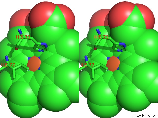
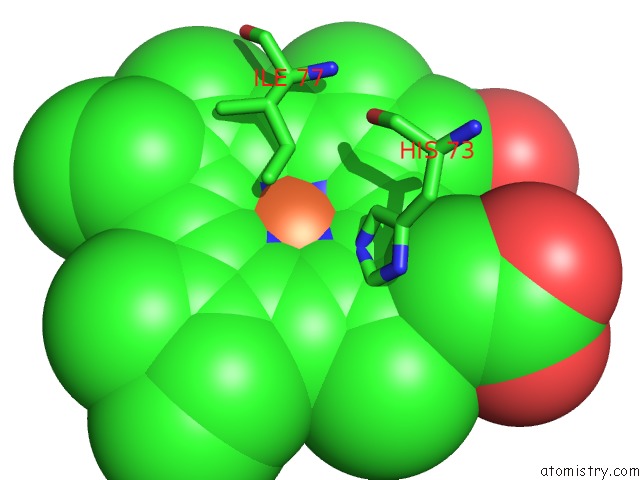
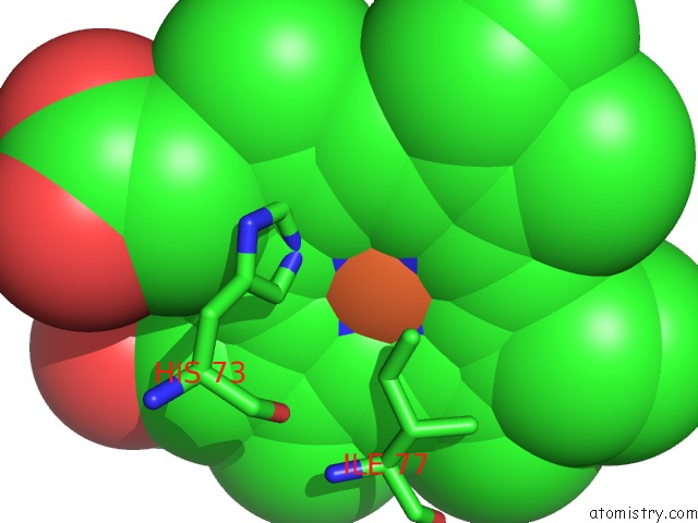
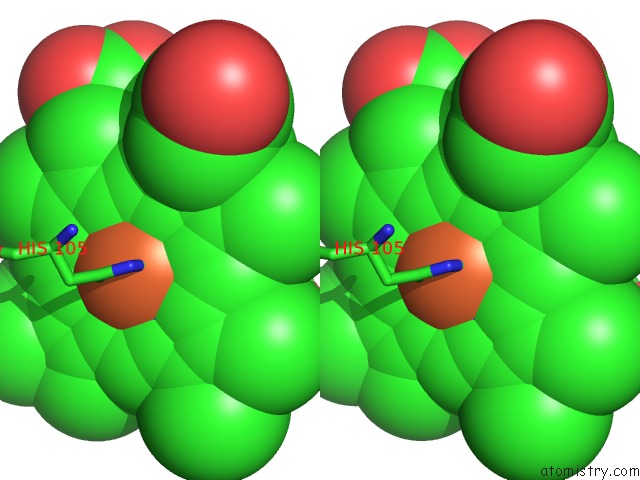
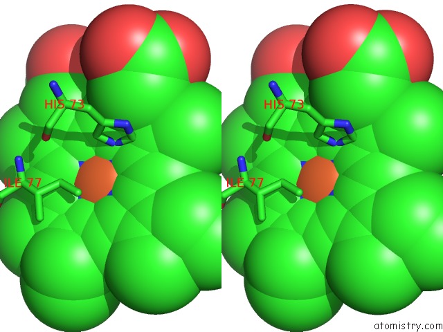
Iron binding site 1 out of 12 in 3lhb
Go back to
Iron binding site 1 out
of 12 in the The 2.7 Angstrom Crystal Structure of Deoxygenated Hemoglobin From the Sea Lamprey (Petromyzon Marinus)
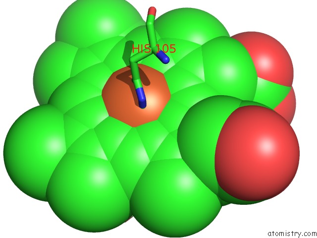
Mono view

Stereo pair view
Mono view
Stereo pair view
A full contact list of Iron with other atoms in the Fe binding
site number 1 of The 2.7 Angstrom Crystal Structure of Deoxygenated Hemoglobin From the Sea Lamprey (Petromyzon Marinus) within 5.0Å range:
|
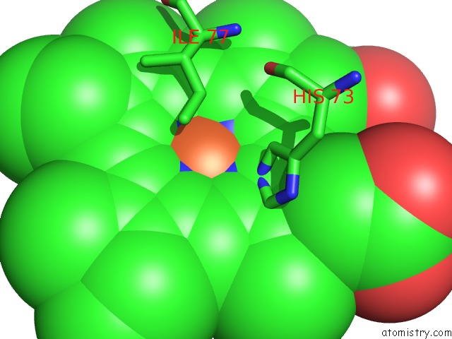
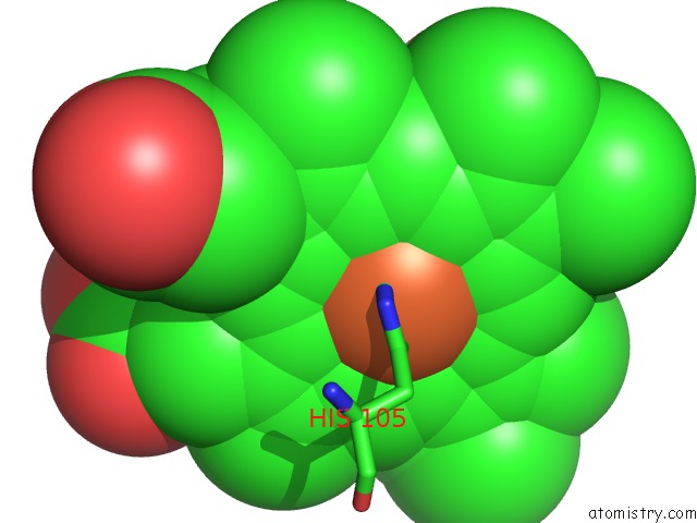
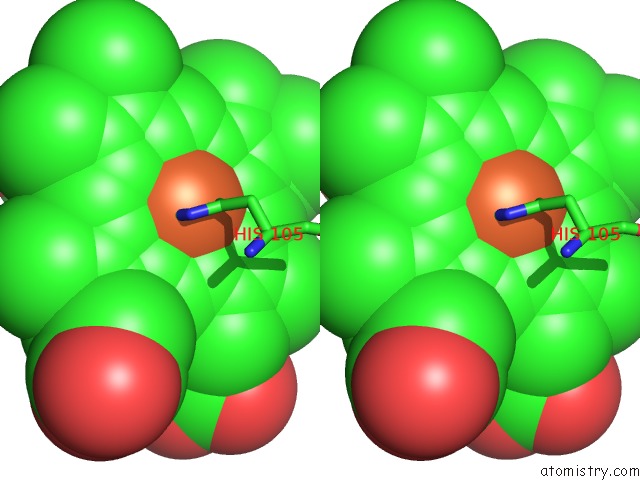
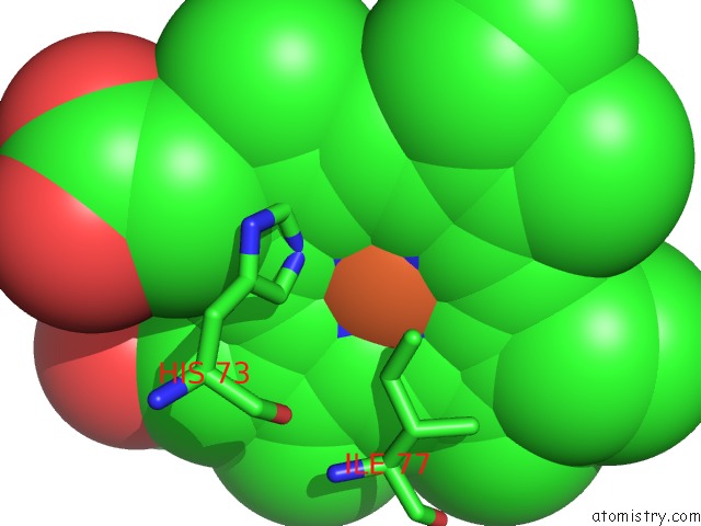
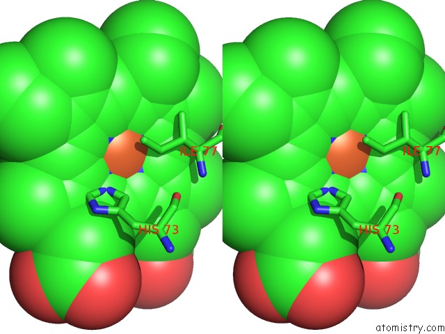
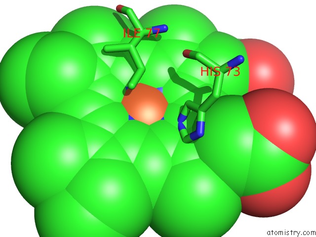
Iron binding site 2 out of 12 in 3lhb
Go back to
Iron binding site 2 out
of 12 in the The 2.7 Angstrom Crystal Structure of Deoxygenated Hemoglobin From the Sea Lamprey (Petromyzon Marinus)
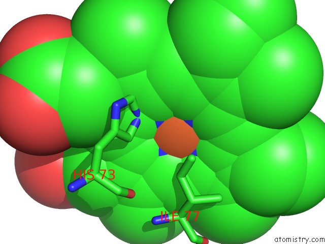
Mono view
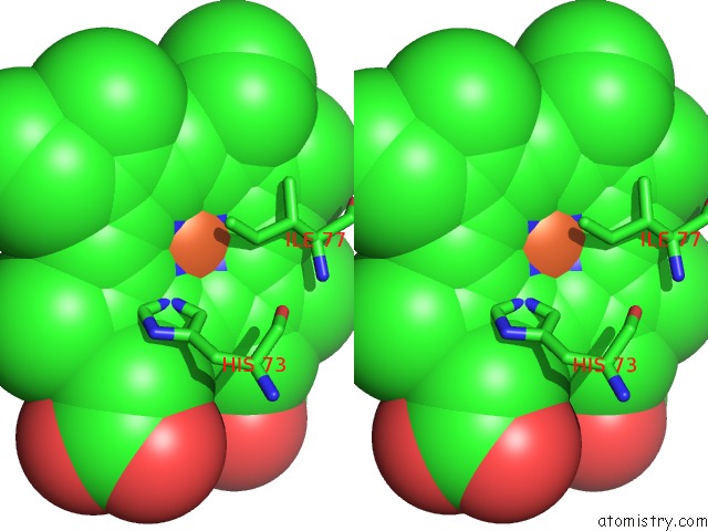
Stereo pair view
Mono view
Stereo pair view
A full contact list of Iron with other atoms in the Fe binding
site number 2 of The 2.7 Angstrom Crystal Structure of Deoxygenated Hemoglobin From the Sea Lamprey (Petromyzon Marinus) within 5.0Å range:
|
Iron binding site 3 out of 12 in 3lhb
Go back to
Iron binding site 3 out
of 12 in the The 2.7 Angstrom Crystal Structure of Deoxygenated Hemoglobin From the Sea Lamprey (Petromyzon Marinus)
Mono view
Stereo pair view
Mono view
Stereo pair view
A full contact list of Iron with other atoms in the Fe binding
site number 3 of The 2.7 Angstrom Crystal Structure of Deoxygenated Hemoglobin From the Sea Lamprey (Petromyzon Marinus) within 5.0Å range:
|
Iron binding site 4 out of 12 in 3lhb
Go back to
Iron binding site 4 out
of 12 in the The 2.7 Angstrom Crystal Structure of Deoxygenated Hemoglobin From the Sea Lamprey (Petromyzon Marinus)
Mono view
Stereo pair view
Mono view
Stereo pair view
A full contact list of Iron with other atoms in the Fe binding
site number 4 of The 2.7 Angstrom Crystal Structure of Deoxygenated Hemoglobin From the Sea Lamprey (Petromyzon Marinus) within 5.0Å range:
|
Iron binding site 5 out of 12 in 3lhb
Go back to
Iron binding site 5 out
of 12 in the The 2.7 Angstrom Crystal Structure of Deoxygenated Hemoglobin From the Sea Lamprey (Petromyzon Marinus)
Mono view

Stereo pair view
Mono view
Stereo pair view
A full contact list of Iron with other atoms in the Fe binding
site number 5 of The 2.7 Angstrom Crystal Structure of Deoxygenated Hemoglobin From the Sea Lamprey (Petromyzon Marinus) within 5.0Å range:
|
Iron binding site 6 out of 12 in 3lhb
Go back to
Iron binding site 6 out
of 12 in the The 2.7 Angstrom Crystal Structure of Deoxygenated Hemoglobin From the Sea Lamprey (Petromyzon Marinus)
Mono view

Stereo pair view
Mono view
Stereo pair view
A full contact list of Iron with other atoms in the Fe binding
site number 6 of The 2.7 Angstrom Crystal Structure of Deoxygenated Hemoglobin From the Sea Lamprey (Petromyzon Marinus) within 5.0Å range:
|
Iron binding site 7 out of 12 in 3lhb
Go back to
Iron binding site 7 out
of 12 in the The 2.7 Angstrom Crystal Structure of Deoxygenated Hemoglobin From the Sea Lamprey (Petromyzon Marinus)
Mono view
Stereo pair view
Mono view
Stereo pair view
A full contact list of Iron with other atoms in the Fe binding
site number 7 of The 2.7 Angstrom Crystal Structure of Deoxygenated Hemoglobin From the Sea Lamprey (Petromyzon Marinus) within 5.0Å range:
|
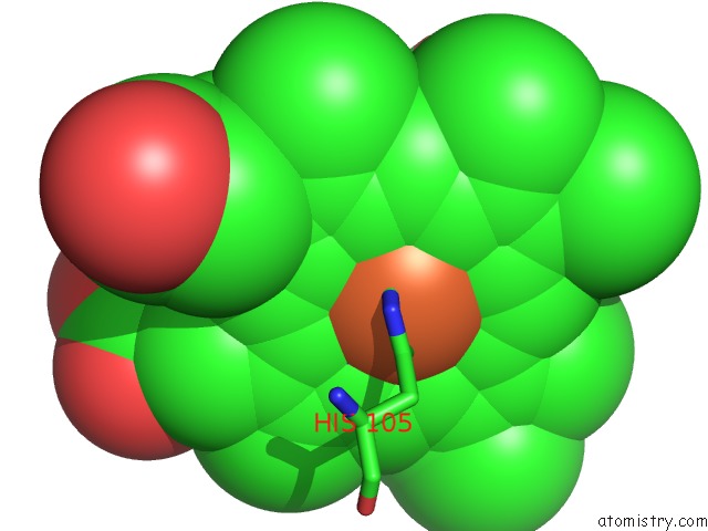
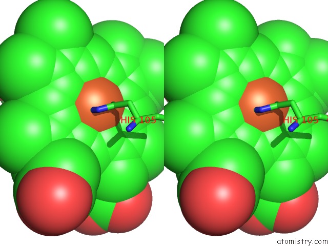
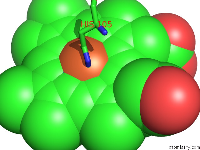
Iron binding site 8 out of 12 in 3lhb
Go back to
Iron binding site 8 out
of 12 in the The 2.7 Angstrom Crystal Structure of Deoxygenated Hemoglobin From the Sea Lamprey (Petromyzon Marinus)
Mono view
Stereo pair view
Mono view
Stereo pair view
A full contact list of Iron with other atoms in the Fe binding
site number 8 of The 2.7 Angstrom Crystal Structure of Deoxygenated Hemoglobin From the Sea Lamprey (Petromyzon Marinus) within 5.0Å range:
|
Iron binding site 9 out of 12 in 3lhb
Go back to
Iron binding site 9 out
of 12 in the The 2.7 Angstrom Crystal Structure of Deoxygenated Hemoglobin From the Sea Lamprey (Petromyzon Marinus)
Mono view
Stereo pair view
Mono view
Stereo pair view
A full contact list of Iron with other atoms in the Fe binding
site number 9 of The 2.7 Angstrom Crystal Structure of Deoxygenated Hemoglobin From the Sea Lamprey (Petromyzon Marinus) within 5.0Å range:
|
Iron binding site 10 out of 12 in 3lhb
Go back to
Iron binding site 10 out
of 12 in the The 2.7 Angstrom Crystal Structure of Deoxygenated Hemoglobin From the Sea Lamprey (Petromyzon Marinus)
Mono view
Stereo pair view
Mono view
Stereo pair view
A full contact list of Iron with other atoms in the Fe binding
site number 10 of The 2.7 Angstrom Crystal Structure of Deoxygenated Hemoglobin From the Sea Lamprey (Petromyzon Marinus) within 5.0Å range:
|
Reference:
H.A.Heaslet,
W.E.Royer Jr..
The 2.7 A Crystal Structure of Deoxygenated Hemoglobin From the Sea Lamprey (Petromyzon Marinus): Structural Basis For A Lowered Oxygen Affinity and Bohr Effect. Structure Fold.Des. V. 7 517 1999.
ISSN: ISSN 0969-2126
PubMed: 10378271
DOI: 10.1016/S0969-2126(99)80068-9
Page generated: Sun Aug 4 14:21:26 2024
ISSN: ISSN 0969-2126
PubMed: 10378271
DOI: 10.1016/S0969-2126(99)80068-9
Last articles
Zn in 9MJ5Zn in 9HNW
Zn in 9G0L
Zn in 9FNE
Zn in 9DZN
Zn in 9E0I
Zn in 9D32
Zn in 9DAK
Zn in 8ZXC
Zn in 8ZUF