Iron »
PDB 3lhs-3m2i »
3ljf »
Iron in PDB 3ljf: The X-Ray Structure of Iron Superoxide Dismutase From Pseudoalteromonas Haloplanktis (Crystal Form II)
Enzymatic activity of The X-Ray Structure of Iron Superoxide Dismutase From Pseudoalteromonas Haloplanktis (Crystal Form II)
All present enzymatic activity of The X-Ray Structure of Iron Superoxide Dismutase From Pseudoalteromonas Haloplanktis (Crystal Form II):
1.15.1.1;
1.15.1.1;
Protein crystallography data
The structure of The X-Ray Structure of Iron Superoxide Dismutase From Pseudoalteromonas Haloplanktis (Crystal Form II), PDB code: 3ljf
was solved by
A.Merlino,
I.Russo Krauss,
B.Rossi,
M.Conte,
A.Vergara,
F.Sica,
with X-Ray Crystallography technique. A brief refinement statistics is given in the table below:
| Resolution Low / High (Å) | 30.00 / 2.10 |
| Space group | P 1 21 1 |
| Cell size a, b, c (Å), α, β, γ (°) | 50.476, 103.785, 90.251, 90.00, 103.81, 90.00 |
| R / Rfree (%) | 19.2 / 24.8 |
Iron Binding Sites:
The binding sites of Iron atom in the The X-Ray Structure of Iron Superoxide Dismutase From Pseudoalteromonas Haloplanktis (Crystal Form II)
(pdb code 3ljf). This binding sites where shown within
5.0 Angstroms radius around Iron atom.
In total 4 binding sites of Iron where determined in the The X-Ray Structure of Iron Superoxide Dismutase From Pseudoalteromonas Haloplanktis (Crystal Form II), PDB code: 3ljf:
Jump to Iron binding site number: 1; 2; 3; 4;
In total 4 binding sites of Iron where determined in the The X-Ray Structure of Iron Superoxide Dismutase From Pseudoalteromonas Haloplanktis (Crystal Form II), PDB code: 3ljf:
Jump to Iron binding site number: 1; 2; 3; 4;
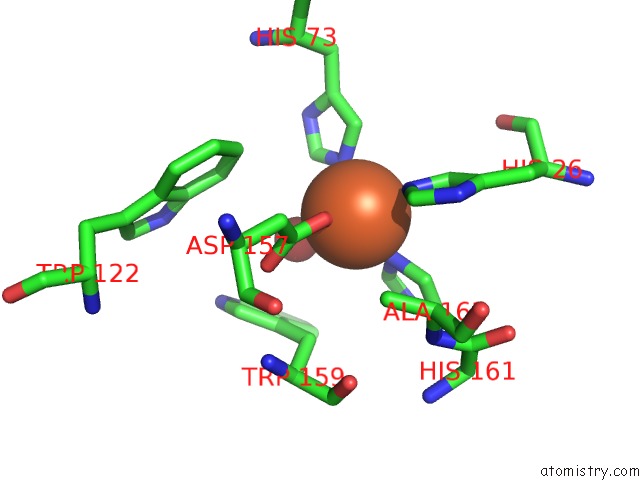
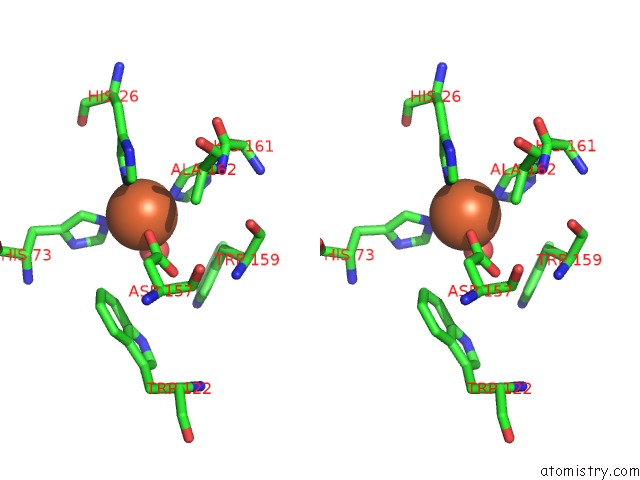
Iron binding site 1 out of 4 in 3ljf
Go back to
Iron binding site 1 out
of 4 in the The X-Ray Structure of Iron Superoxide Dismutase From Pseudoalteromonas Haloplanktis (Crystal Form II)

Mono view
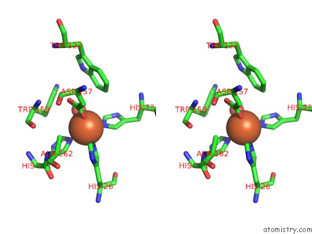
Stereo pair view
Mono view
Stereo pair view
A full contact list of Iron with other atoms in the Fe binding
site number 1 of The X-Ray Structure of Iron Superoxide Dismutase From Pseudoalteromonas Haloplanktis (Crystal Form II) within 5.0Å range:
|
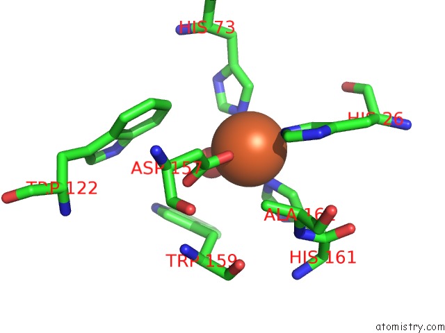
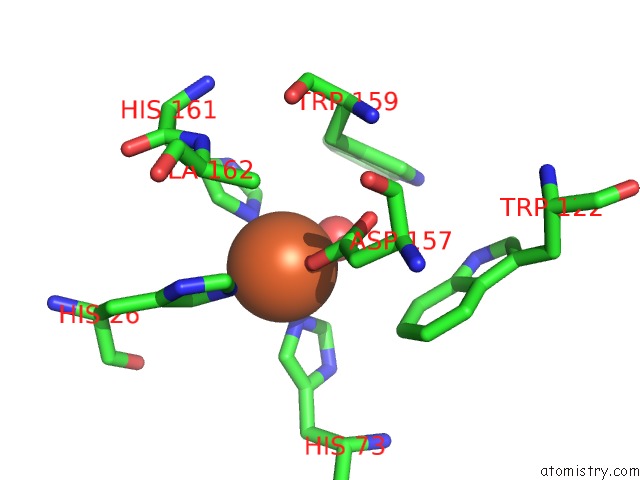
Iron binding site 2 out of 4 in 3ljf
Go back to
Iron binding site 2 out
of 4 in the The X-Ray Structure of Iron Superoxide Dismutase From Pseudoalteromonas Haloplanktis (Crystal Form II)
Mono view
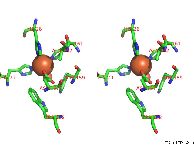
Stereo pair view
Mono view
Stereo pair view
A full contact list of Iron with other atoms in the Fe binding
site number 2 of The X-Ray Structure of Iron Superoxide Dismutase From Pseudoalteromonas Haloplanktis (Crystal Form II) within 5.0Å range:
|
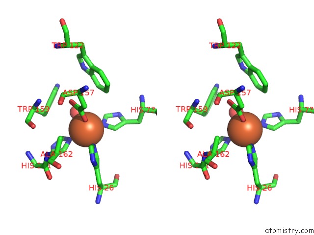
Iron binding site 3 out of 4 in 3ljf
Go back to
Iron binding site 3 out
of 4 in the The X-Ray Structure of Iron Superoxide Dismutase From Pseudoalteromonas Haloplanktis (Crystal Form II)
Mono view
Stereo pair view
Mono view
Stereo pair view
A full contact list of Iron with other atoms in the Fe binding
site number 3 of The X-Ray Structure of Iron Superoxide Dismutase From Pseudoalteromonas Haloplanktis (Crystal Form II) within 5.0Å range:
|
Iron binding site 4 out of 4 in 3ljf
Go back to
Iron binding site 4 out
of 4 in the The X-Ray Structure of Iron Superoxide Dismutase From Pseudoalteromonas Haloplanktis (Crystal Form II)
Mono view
Stereo pair view
Mono view
Stereo pair view
A full contact list of Iron with other atoms in the Fe binding
site number 4 of The X-Ray Structure of Iron Superoxide Dismutase From Pseudoalteromonas Haloplanktis (Crystal Form II) within 5.0Å range:
|
Reference:
A.Merlino,
I.Russo Krauss,
I.Castellano,
E.De Vendittis,
B.Rossi,
M.Conte,
A.Vergara,
F.Sica.
Structure and Flexibility in Cold-Adapted Iron Superoxide Dismutases: the Case of the Enzyme Isolated From Pseudoalteromonas Haloplanktis. J.Struct.Biol. V. 172 343 2010.
ISSN: ISSN 1047-8477
PubMed: 20732427
DOI: 10.1016/J.JSB.2010.08.008
Page generated: Sun Aug 4 14:30:19 2024
ISSN: ISSN 1047-8477
PubMed: 20732427
DOI: 10.1016/J.JSB.2010.08.008
Last articles
F in 7QM8F in 7QM7
F in 7QM5
F in 7QM4
F in 7QLT
F in 7QIT
F in 7QM3
F in 7QH1
F in 7QIS
F in 7QK4