Iron »
PDB 3lhs-3m2i »
3lr9 »
Iron in PDB 3lr9: X-Ray Photogenerated Ferrous Horse Heart Myoglobin, Nitrite Adduct
Protein crystallography data
The structure of X-Ray Photogenerated Ferrous Horse Heart Myoglobin, Nitrite Adduct, PDB code: 3lr9
was solved by
J.Yi,
A.M.Orville,
G.B.Richter-Addo,
with X-Ray Crystallography technique. A brief refinement statistics is given in the table below:
| Resolution Low / High (Å) | 26.74 / 1.55 |
| Space group | P 1 21 1 |
| Cell size a, b, c (Å), α, β, γ (°) | 35.327, 28.691, 62.911, 90.00, 105.64, 90.00 |
| R / Rfree (%) | 17.8 / 21.2 |
Iron Binding Sites:
The binding sites of Iron atom in the X-Ray Photogenerated Ferrous Horse Heart Myoglobin, Nitrite Adduct
(pdb code 3lr9). This binding sites where shown within
5.0 Angstroms radius around Iron atom.
In total only one binding site of Iron was determined in the X-Ray Photogenerated Ferrous Horse Heart Myoglobin, Nitrite Adduct, PDB code: 3lr9:
In total only one binding site of Iron was determined in the X-Ray Photogenerated Ferrous Horse Heart Myoglobin, Nitrite Adduct, PDB code: 3lr9:
Iron binding site 1 out of 1 in 3lr9
Go back to
Iron binding site 1 out
of 1 in the X-Ray Photogenerated Ferrous Horse Heart Myoglobin, Nitrite Adduct
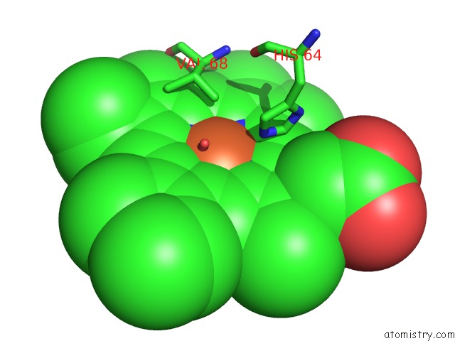
Mono view
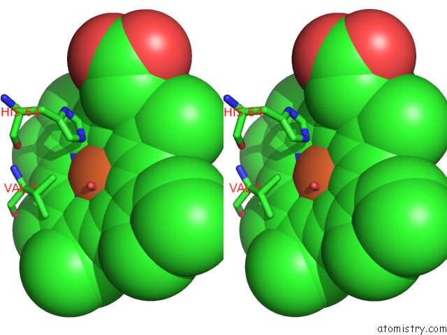
Stereo pair view
Mono view
Stereo pair view
A full contact list of Iron with other atoms in the Fe binding
site number 1 of X-Ray Photogenerated Ferrous Horse Heart Myoglobin, Nitrite Adduct within 5.0Å range:
|
Reference:
J.Yi,
A.M.Orville,
J.M.Skinner,
M.J.Skinner,
G.B.Richter-Addo.
Synchrotron X-Ray-Induced Photoreduction of Ferric Myoglobin Nitrite Crystals Gives the Ferrous Derivative with Retention of the O-Bonded Nitrite Ligand. Biochemistry V. 49 5969 2010.
ISSN: ISSN 0006-2960
PubMed: 20568729
DOI: 10.1021/BI100801G
Page generated: Sun Aug 4 14:32:44 2024
ISSN: ISSN 0006-2960
PubMed: 20568729
DOI: 10.1021/BI100801G
Last articles
Zn in 9JYWZn in 9IR4
Zn in 9IR3
Zn in 9GMX
Zn in 9GMW
Zn in 9JEJ
Zn in 9ERF
Zn in 9ERE
Zn in 9EGV
Zn in 9EGW