Iron »
PDB 3m38-3mma »
3m5q »
Iron in PDB 3m5q: 0.93 A Structure of Manganese-Bound Manganese Peroxidase
Enzymatic activity of 0.93 A Structure of Manganese-Bound Manganese Peroxidase
All present enzymatic activity of 0.93 A Structure of Manganese-Bound Manganese Peroxidase:
1.11.1.13;
1.11.1.13;
Protein crystallography data
The structure of 0.93 A Structure of Manganese-Bound Manganese Peroxidase, PDB code: 3m5q
was solved by
M.Sundaramoorthy,
M.H.Gold,
T.L.Poulos,
with X-Ray Crystallography technique. A brief refinement statistics is given in the table below:
| Resolution Low / High (Å) | 8.00 / 0.93 |
| Space group | C 1 2 1 |
| Cell size a, b, c (Å), α, β, γ (°) | 160.570, 45.300, 52.830, 90.00, 97.31, 90.00 |
| R / Rfree (%) | n/a / 13.4 |
Other elements in 3m5q:
The structure of 0.93 A Structure of Manganese-Bound Manganese Peroxidase also contains other interesting chemical elements:
| Manganese | (Mn) | 1 atom |
| Calcium | (Ca) | 2 atoms |
Iron Binding Sites:
The binding sites of Iron atom in the 0.93 A Structure of Manganese-Bound Manganese Peroxidase
(pdb code 3m5q). This binding sites where shown within
5.0 Angstroms radius around Iron atom.
In total only one binding site of Iron was determined in the 0.93 A Structure of Manganese-Bound Manganese Peroxidase, PDB code: 3m5q:
In total only one binding site of Iron was determined in the 0.93 A Structure of Manganese-Bound Manganese Peroxidase, PDB code: 3m5q:
Iron binding site 1 out of 1 in 3m5q
Go back to
Iron binding site 1 out
of 1 in the 0.93 A Structure of Manganese-Bound Manganese Peroxidase
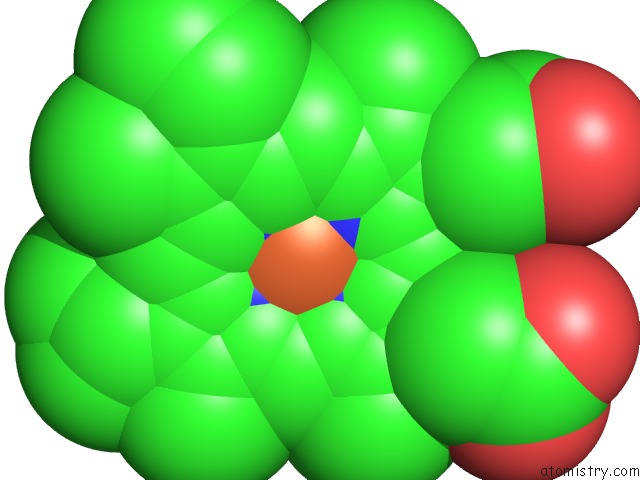
Mono view
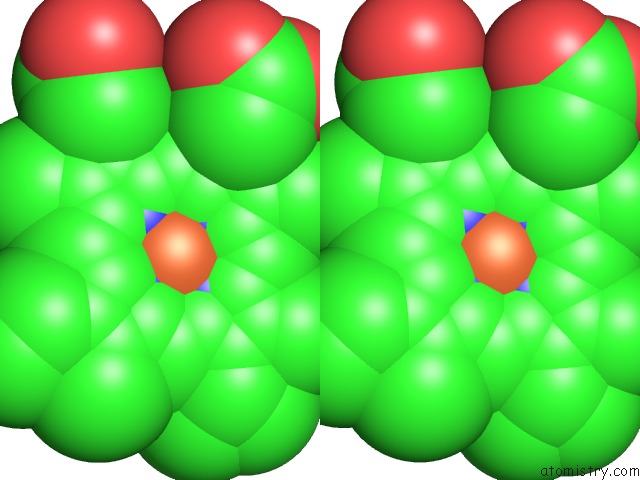
Stereo pair view
Mono view
Stereo pair view
A full contact list of Iron with other atoms in the Fe binding
site number 1 of 0.93 A Structure of Manganese-Bound Manganese Peroxidase within 5.0Å range:
|
Reference:
M.Sundaramoorthy,
M.H.Gold,
T.L.Poulos.
Ultrahigh (0.93A) Resolution Structure of Manganese Peroxidase From Phanerochaete Chrysosporium: Implications For the Catalytic Mechanism. J.Inorg.Biochem. V. 104 683 2010.
ISSN: ISSN 0162-0134
PubMed: 20356630
DOI: 10.1016/J.JINORGBIO.2010.02.011
Page generated: Sun Aug 4 14:43:54 2024
ISSN: ISSN 0162-0134
PubMed: 20356630
DOI: 10.1016/J.JINORGBIO.2010.02.011
Last articles
Zn in 9J0NZn in 9J0O
Zn in 9J0P
Zn in 9FJX
Zn in 9EKB
Zn in 9C0F
Zn in 9CAH
Zn in 9CH0
Zn in 9CH3
Zn in 9CH1