Iron »
PDB 3m38-3mma »
3mdm »
Iron in PDB 3mdm: Thioperamide Complex of Cytochrome P450 46A1
Enzymatic activity of Thioperamide Complex of Cytochrome P450 46A1
All present enzymatic activity of Thioperamide Complex of Cytochrome P450 46A1:
1.14.13.98;
1.14.13.98;
Protein crystallography data
The structure of Thioperamide Complex of Cytochrome P450 46A1, PDB code: 3mdm
was solved by
N.Mast,
C.Charvet,
I.Pikuleva,
C.D.Stout,
with X-Ray Crystallography technique. A brief refinement statistics is given in the table below:
| Resolution Low / High (Å) | 20.00 / 1.60 |
| Space group | P 21 21 21 |
| Cell size a, b, c (Å), α, β, γ (°) | 58.830, 85.820, 104.200, 90.00, 90.00, 90.00 |
| R / Rfree (%) | 18.2 / 22.6 |
Iron Binding Sites:
The binding sites of Iron atom in the Thioperamide Complex of Cytochrome P450 46A1
(pdb code 3mdm). This binding sites where shown within
5.0 Angstroms radius around Iron atom.
In total only one binding site of Iron was determined in the Thioperamide Complex of Cytochrome P450 46A1, PDB code: 3mdm:
In total only one binding site of Iron was determined in the Thioperamide Complex of Cytochrome P450 46A1, PDB code: 3mdm:
Iron binding site 1 out of 1 in 3mdm
Go back to
Iron binding site 1 out
of 1 in the Thioperamide Complex of Cytochrome P450 46A1
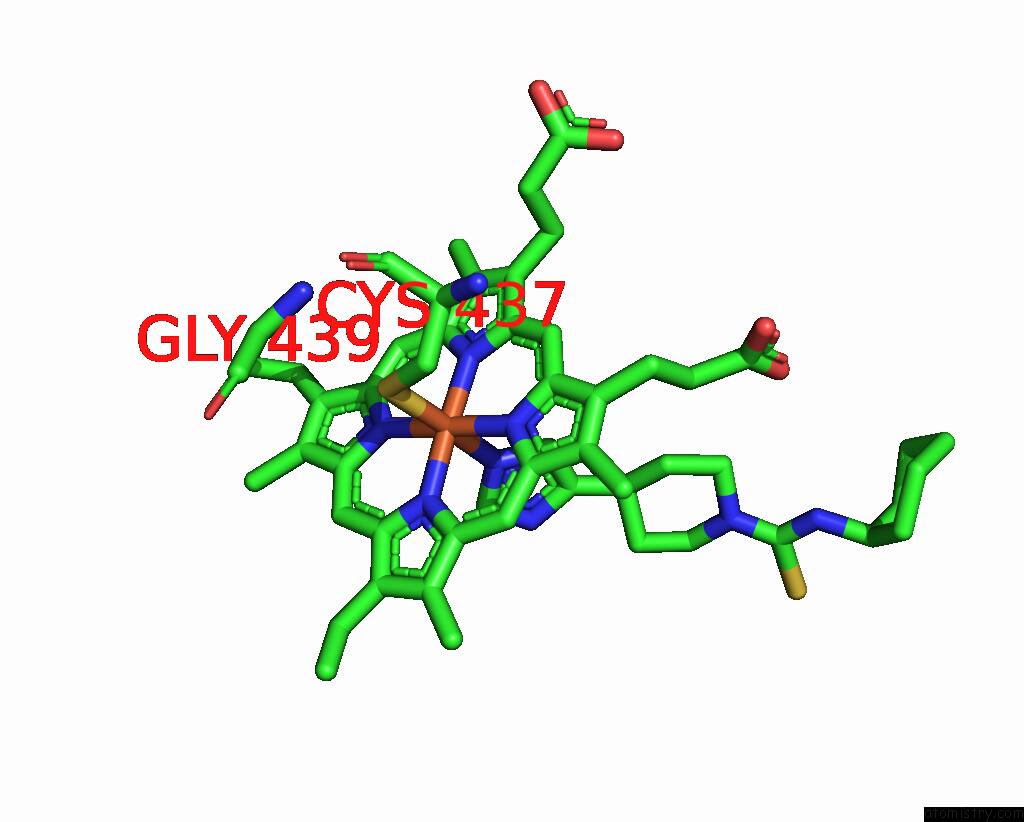
Mono view
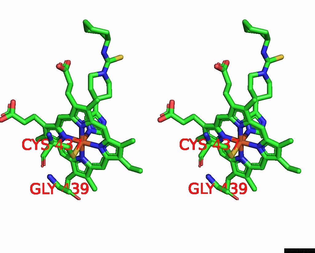
Stereo pair view
Mono view
Stereo pair view
A full contact list of Iron with other atoms in the Fe binding
site number 1 of Thioperamide Complex of Cytochrome P450 46A1 within 5.0Å range:
|
Reference:
N.Mast,
C.Charvet,
I.A.Pikuleva,
C.D.Stout.
Structural Basis of Drug Binding to CYP46A1, An Enzyme That Controls Cholesterol Turnover in the Brain. J.Biol.Chem. V. 285 31783 2010.
ISSN: ISSN 0021-9258
PubMed: 20667828
DOI: 10.1074/JBC.M110.143313
Page generated: Sun Aug 4 14:45:22 2024
ISSN: ISSN 0021-9258
PubMed: 20667828
DOI: 10.1074/JBC.M110.143313
Last articles
Fe in 2YXOFe in 2YRS
Fe in 2YXC
Fe in 2YNM
Fe in 2YVJ
Fe in 2YP1
Fe in 2YU2
Fe in 2YU1
Fe in 2YQB
Fe in 2YOO