Iron »
PDB 3m38-3mma »
3mgu »
Iron in PDB 3mgu: Structure of S. Cerevisiae TPA1 Protein, A Proline Hydroxylase Modifying Ribosomal Protein RPS23
Protein crystallography data
The structure of Structure of S. Cerevisiae TPA1 Protein, A Proline Hydroxylase Modifying Ribosomal Protein RPS23, PDB code: 3mgu
was solved by
J.Henri,
D.Rispal,
E.Bayart,
H.Van Tilbeurgh,
B.Seraphin,
M.Graille,
with X-Ray Crystallography technique. A brief refinement statistics is given in the table below:
| Resolution Low / High (Å) | 19.92 / 2.80 |
| Space group | C 2 2 21 |
| Cell size a, b, c (Å), α, β, γ (°) | 81.180, 104.840, 205.570, 90.00, 90.00, 90.00 |
| R / Rfree (%) | 23.9 / 29.6 |
Iron Binding Sites:
The binding sites of Iron atom in the Structure of S. Cerevisiae TPA1 Protein, A Proline Hydroxylase Modifying Ribosomal Protein RPS23
(pdb code 3mgu). This binding sites where shown within
5.0 Angstroms radius around Iron atom.
In total only one binding site of Iron was determined in the Structure of S. Cerevisiae TPA1 Protein, A Proline Hydroxylase Modifying Ribosomal Protein RPS23, PDB code: 3mgu:
In total only one binding site of Iron was determined in the Structure of S. Cerevisiae TPA1 Protein, A Proline Hydroxylase Modifying Ribosomal Protein RPS23, PDB code: 3mgu:
Iron binding site 1 out of 1 in 3mgu
Go back to
Iron binding site 1 out
of 1 in the Structure of S. Cerevisiae TPA1 Protein, A Proline Hydroxylase Modifying Ribosomal Protein RPS23
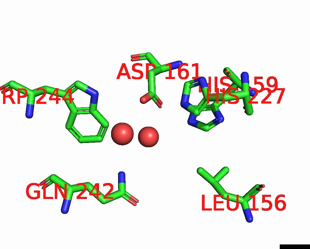
Mono view
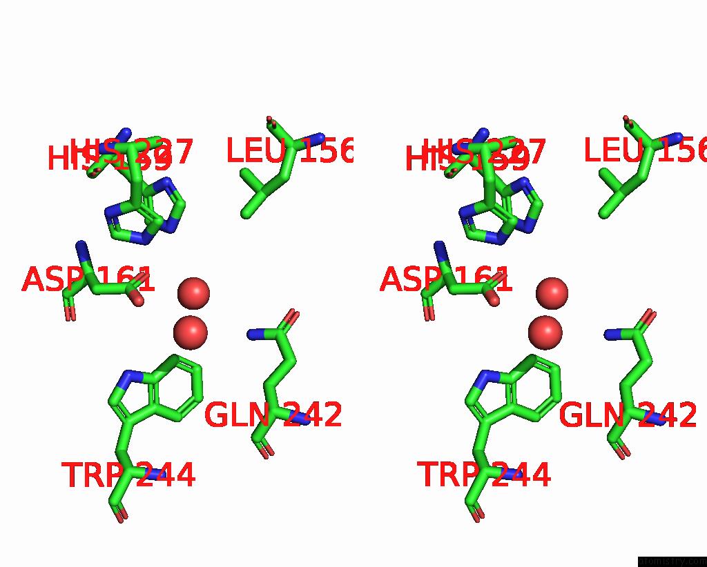
Stereo pair view
Mono view
Stereo pair view
A full contact list of Iron with other atoms in the Fe binding
site number 1 of Structure of S. Cerevisiae TPA1 Protein, A Proline Hydroxylase Modifying Ribosomal Protein RPS23 within 5.0Å range:
|
Reference:
J.Henri,
D.Rispal,
E.Bayart,
H.Van Tilbeurgh,
B.Seraphin,
M.Graille.
Structural and Functional Insights Into S. Cerevisiae TPA1, A Putative Prolyl Hydroxylase Influencing Translation Termination and Transcription To Be Published.
Page generated: Tue Aug 5 03:46:49 2025
Last articles
Fe in 3UNIFe in 3UNC
Fe in 3UNA
Fe in 3UI0
Fe in 3UHZ
Fe in 3UK4
Fe in 3UHY
Fe in 3UHX
Fe in 3UHW
Fe in 3UHV