Iron »
PDB 3mmb-3n5r »
3mom »
Iron in PDB 3mom: Structure of Holo Hasap H32A Mutant Complexed with Imidazole From Pseudomonas Aeruginosa to 2.25A Resolution
Protein crystallography data
The structure of Structure of Holo Hasap H32A Mutant Complexed with Imidazole From Pseudomonas Aeruginosa to 2.25A Resolution, PDB code: 3mom
was solved by
S.Lovell,
K.P.Battaile,
G.Jepkorir,
J.C.Rodriguez,
H.Rui,
W.Im,
A.Y.Alontaga,
E.Yukl,
P.Moenne-Loccoz,
M.Rivera,
with X-Ray Crystallography technique. A brief refinement statistics is given in the table below:
| Resolution Low / High (Å) | 24.64 / 2.25 |
| Space group | P 1 2 1 |
| Cell size a, b, c (Å), α, β, γ (°) | 47.373, 46.378, 81.088, 90.00, 97.10, 90.00 |
| R / Rfree (%) | 21.4 / 26.7 |
Iron Binding Sites:
The binding sites of Iron atom in the Structure of Holo Hasap H32A Mutant Complexed with Imidazole From Pseudomonas Aeruginosa to 2.25A Resolution
(pdb code 3mom). This binding sites where shown within
5.0 Angstroms radius around Iron atom.
In total 2 binding sites of Iron where determined in the Structure of Holo Hasap H32A Mutant Complexed with Imidazole From Pseudomonas Aeruginosa to 2.25A Resolution, PDB code: 3mom:
Jump to Iron binding site number: 1; 2;
In total 2 binding sites of Iron where determined in the Structure of Holo Hasap H32A Mutant Complexed with Imidazole From Pseudomonas Aeruginosa to 2.25A Resolution, PDB code: 3mom:
Jump to Iron binding site number: 1; 2;
Iron binding site 1 out of 2 in 3mom
Go back to
Iron binding site 1 out
of 2 in the Structure of Holo Hasap H32A Mutant Complexed with Imidazole From Pseudomonas Aeruginosa to 2.25A Resolution
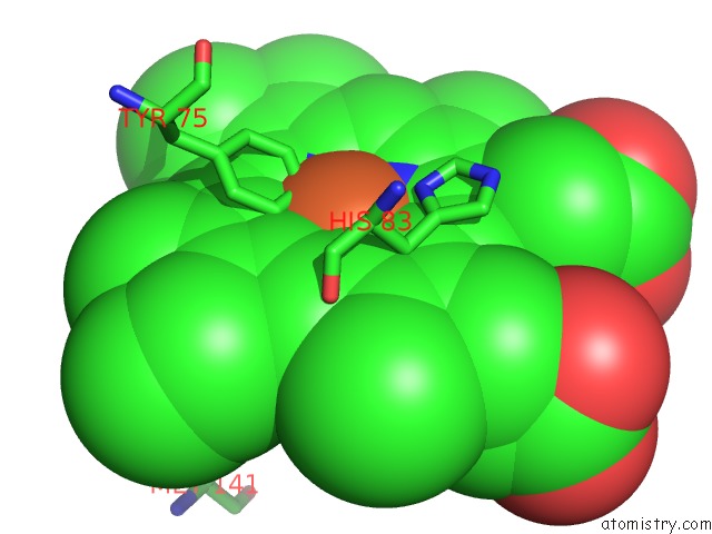
Mono view
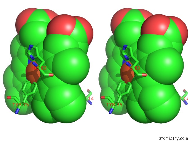
Stereo pair view
Mono view
Stereo pair view
A full contact list of Iron with other atoms in the Fe binding
site number 1 of Structure of Holo Hasap H32A Mutant Complexed with Imidazole From Pseudomonas Aeruginosa to 2.25A Resolution within 5.0Å range:
|
Iron binding site 2 out of 2 in 3mom
Go back to
Iron binding site 2 out
of 2 in the Structure of Holo Hasap H32A Mutant Complexed with Imidazole From Pseudomonas Aeruginosa to 2.25A Resolution
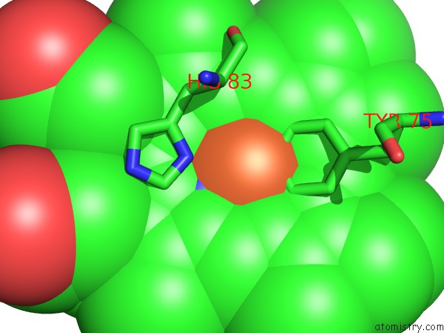
Mono view
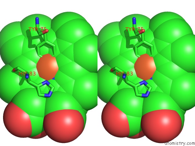
Stereo pair view
Mono view
Stereo pair view
A full contact list of Iron with other atoms in the Fe binding
site number 2 of Structure of Holo Hasap H32A Mutant Complexed with Imidazole From Pseudomonas Aeruginosa to 2.25A Resolution within 5.0Å range:
|
Reference:
G.Jepkorir,
J.C.Rodriguez,
H.Rui,
W.Im,
S.Lovell,
K.P.Battaile,
A.Y.Alontaga,
E.T.Yukl,
M.Rivera.
Structural, uc(Nmr) Spectroscopic, and Computational Investigation of Hemin Loading in the Hemophore Hasap From Pseudomonas Aeruginosa. J.Am.Chem.Soc. V. 132 9857 2010.
ISSN: ISSN 0002-7863
PubMed: 20572666
DOI: 10.1021/JA103498Z
Page generated: Sun Aug 4 15:40:17 2024
ISSN: ISSN 0002-7863
PubMed: 20572666
DOI: 10.1021/JA103498Z
Last articles
Zn in 9MJ5Zn in 9HNW
Zn in 9G0L
Zn in 9FNE
Zn in 9DZN
Zn in 9E0I
Zn in 9D32
Zn in 9DAK
Zn in 8ZXC
Zn in 8ZUF