Iron »
PDB 3mmb-3n5r »
3mvf »
Iron in PDB 3mvf: Crystal Structure of Nitrophorin 4 From Rhodnius Prolixus Complexed with Nitrite at pH 7.4
Protein crystallography data
The structure of Crystal Structure of Nitrophorin 4 From Rhodnius Prolixus Complexed with Nitrite at pH 7.4, PDB code: 3mvf
was solved by
H.Ogata,
C.He,
M.Knipp,
with X-Ray Crystallography technique. A brief refinement statistics is given in the table below:
| Resolution Low / High (Å) | 15.00 / 1.40 |
| Space group | C 1 2 1 |
| Cell size a, b, c (Å), α, β, γ (°) | 70.301, 42.909, 52.460, 90.00, 94.05, 90.00 |
| R / Rfree (%) | 13.6 / 18.7 |
Iron Binding Sites:
The binding sites of Iron atom in the Crystal Structure of Nitrophorin 4 From Rhodnius Prolixus Complexed with Nitrite at pH 7.4
(pdb code 3mvf). This binding sites where shown within
5.0 Angstroms radius around Iron atom.
In total only one binding site of Iron was determined in the Crystal Structure of Nitrophorin 4 From Rhodnius Prolixus Complexed with Nitrite at pH 7.4, PDB code: 3mvf:
In total only one binding site of Iron was determined in the Crystal Structure of Nitrophorin 4 From Rhodnius Prolixus Complexed with Nitrite at pH 7.4, PDB code: 3mvf:
Iron binding site 1 out of 1 in 3mvf
Go back to
Iron binding site 1 out
of 1 in the Crystal Structure of Nitrophorin 4 From Rhodnius Prolixus Complexed with Nitrite at pH 7.4
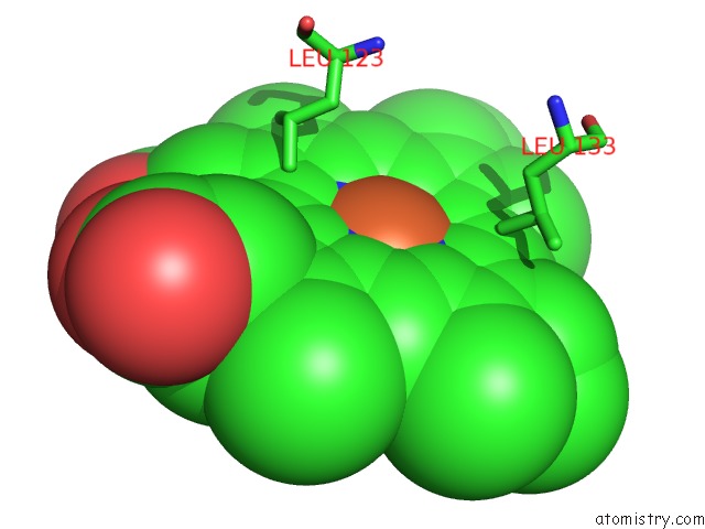
Mono view
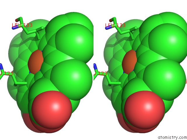
Stereo pair view
Mono view
Stereo pair view
A full contact list of Iron with other atoms in the Fe binding
site number 1 of Crystal Structure of Nitrophorin 4 From Rhodnius Prolixus Complexed with Nitrite at pH 7.4 within 5.0Å range:
|
Reference:
C.He,
H.Ogata,
M.Knipp.
Formation of the Complex of Nitrite with the Ferriheme B Beta-Barrel Proteins Nitrophorin 4 and Nitrophorin 7. Biochemistry V. 49 5841 2010.
ISSN: ISSN 0006-2960
PubMed: 20524697
DOI: 10.1021/BI100324Z
Page generated: Sun Aug 4 15:43:15 2024
ISSN: ISSN 0006-2960
PubMed: 20524697
DOI: 10.1021/BI100324Z
Last articles
Zn in 9MJ5Zn in 9HNW
Zn in 9G0L
Zn in 9FNE
Zn in 9DZN
Zn in 9E0I
Zn in 9D32
Zn in 9DAK
Zn in 8ZXC
Zn in 8ZUF