Iron »
PDB 3mmb-3n5r »
3mvr »
Iron in PDB 3mvr: Crystal Structure of Cytochrome P450 2B4-H226Y in A Closed Conformation
Enzymatic activity of Crystal Structure of Cytochrome P450 2B4-H226Y in A Closed Conformation
All present enzymatic activity of Crystal Structure of Cytochrome P450 2B4-H226Y in A Closed Conformation:
1.14.14.1;
1.14.14.1;
Protein crystallography data
The structure of Crystal Structure of Cytochrome P450 2B4-H226Y in A Closed Conformation, PDB code: 3mvr
was solved by
M.B.Shah,
C.D.Stout,
J.R.Halpert,
with X-Ray Crystallography technique. A brief refinement statistics is given in the table below:
| Resolution Low / High (Å) | 150.00 / 1.76 |
| Space group | P 31 |
| Cell size a, b, c (Å), α, β, γ (°) | 91.470, 91.470, 150.400, 90.00, 90.00, 120.00 |
| R / Rfree (%) | 19.6 / 22.4 |
Iron Binding Sites:
The binding sites of Iron atom in the Crystal Structure of Cytochrome P450 2B4-H226Y in A Closed Conformation
(pdb code 3mvr). This binding sites where shown within
5.0 Angstroms radius around Iron atom.
In total 2 binding sites of Iron where determined in the Crystal Structure of Cytochrome P450 2B4-H226Y in A Closed Conformation, PDB code: 3mvr:
Jump to Iron binding site number: 1; 2;
In total 2 binding sites of Iron where determined in the Crystal Structure of Cytochrome P450 2B4-H226Y in A Closed Conformation, PDB code: 3mvr:
Jump to Iron binding site number: 1; 2;
Iron binding site 1 out of 2 in 3mvr
Go back to
Iron binding site 1 out
of 2 in the Crystal Structure of Cytochrome P450 2B4-H226Y in A Closed Conformation
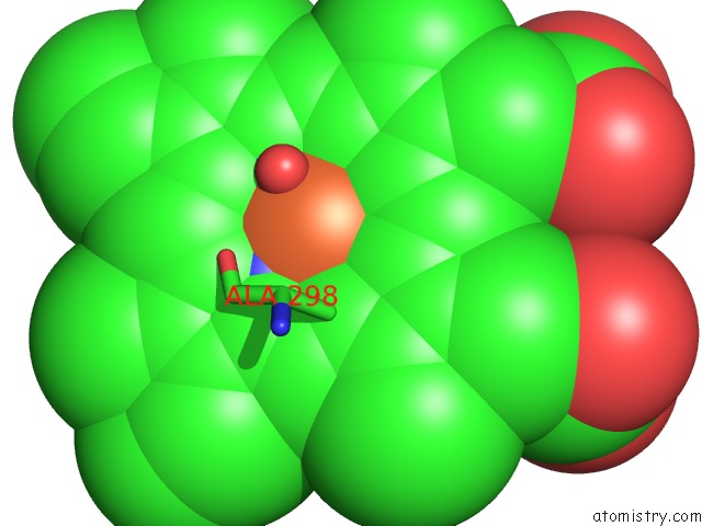
Mono view
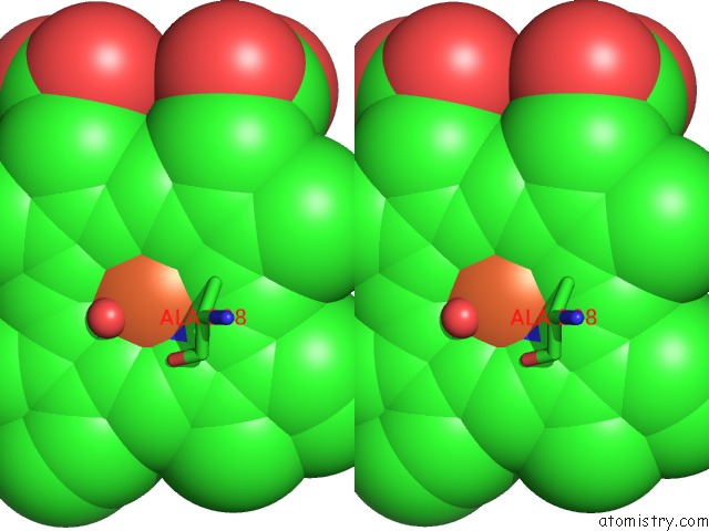
Stereo pair view
Mono view
Stereo pair view
A full contact list of Iron with other atoms in the Fe binding
site number 1 of Crystal Structure of Cytochrome P450 2B4-H226Y in A Closed Conformation within 5.0Å range:
|
Iron binding site 2 out of 2 in 3mvr
Go back to
Iron binding site 2 out
of 2 in the Crystal Structure of Cytochrome P450 2B4-H226Y in A Closed Conformation
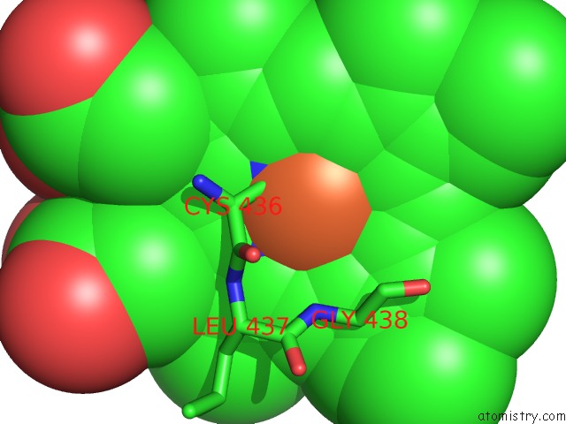
Mono view
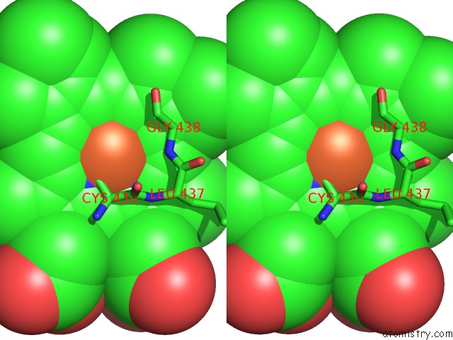
Stereo pair view
Mono view
Stereo pair view
A full contact list of Iron with other atoms in the Fe binding
site number 2 of Crystal Structure of Cytochrome P450 2B4-H226Y in A Closed Conformation within 5.0Å range:
|
Reference:
P.R.Wilderman,
M.B.Shah,
T.Liu,
S.Li,
S.Hsu,
A.G.Roberts,
D.R.Goodlett,
Q.Zhang,
V.L.Woods,
C.D.Stout,
J.R.Halpert.
Plasticity of Cytochrome P450 2B4 As Investigated By Hydrogen-Deuterium Exchange Mass Spectrometry and X-Ray Crystallography. J.Biol.Chem. V. 285 38602 2010.
ISSN: ISSN 0021-9258
PubMed: 20880847
DOI: 10.1074/JBC.M110.180646
Page generated: Sun Aug 4 15:43:21 2024
ISSN: ISSN 0021-9258
PubMed: 20880847
DOI: 10.1074/JBC.M110.180646
Last articles
Zn in 9MJ5Zn in 9HNW
Zn in 9G0L
Zn in 9FNE
Zn in 9DZN
Zn in 9E0I
Zn in 9D32
Zn in 9DAK
Zn in 8ZXC
Zn in 8ZUF