Iron »
PDB 3mmb-3n5r »
3mzs »
Iron in PDB 3mzs: Crystal Structure of Cytochrome P450 CYP11A1 in Complex with 22- Hydroxy-Cholesterol
Enzymatic activity of Crystal Structure of Cytochrome P450 CYP11A1 in Complex with 22- Hydroxy-Cholesterol
All present enzymatic activity of Crystal Structure of Cytochrome P450 CYP11A1 in Complex with 22- Hydroxy-Cholesterol:
1.14.15.6;
1.14.15.6;
Protein crystallography data
The structure of Crystal Structure of Cytochrome P450 CYP11A1 in Complex with 22- Hydroxy-Cholesterol, PDB code: 3mzs
was solved by
C.D.Stout,
A.Annalora,
N.Mast,
I.Pikuleva,
with X-Ray Crystallography technique. A brief refinement statistics is given in the table below:
| Resolution Low / High (Å) | 60.01 / 2.50 |
| Space group | P 1 21 1 |
| Cell size a, b, c (Å), α, β, γ (°) | 109.450, 94.630, 113.500, 90.00, 89.96, 90.00 |
| R / Rfree (%) | 26.6 / 28.1 |
Iron Binding Sites:
The binding sites of Iron atom in the Crystal Structure of Cytochrome P450 CYP11A1 in Complex with 22- Hydroxy-Cholesterol
(pdb code 3mzs). This binding sites where shown within
5.0 Angstroms radius around Iron atom.
In total 4 binding sites of Iron where determined in the Crystal Structure of Cytochrome P450 CYP11A1 in Complex with 22- Hydroxy-Cholesterol, PDB code: 3mzs:
Jump to Iron binding site number: 1; 2; 3; 4;
In total 4 binding sites of Iron where determined in the Crystal Structure of Cytochrome P450 CYP11A1 in Complex with 22- Hydroxy-Cholesterol, PDB code: 3mzs:
Jump to Iron binding site number: 1; 2; 3; 4;
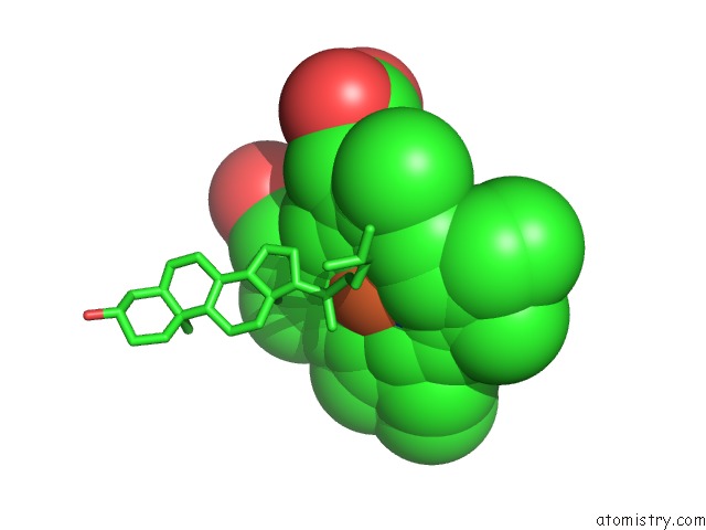
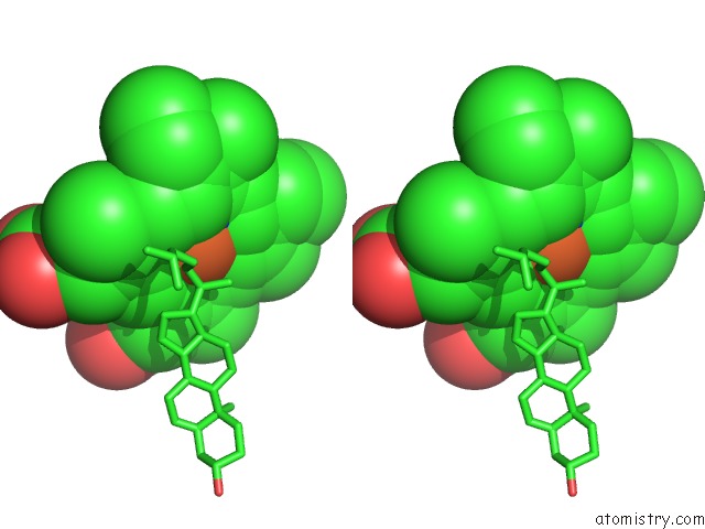
Iron binding site 1 out of 4 in 3mzs
Go back to
Iron binding site 1 out
of 4 in the Crystal Structure of Cytochrome P450 CYP11A1 in Complex with 22- Hydroxy-Cholesterol
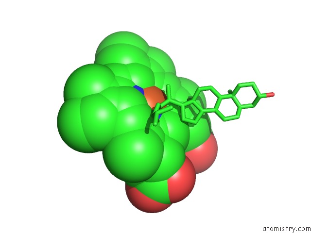
Mono view
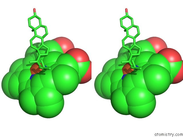
Stereo pair view
Mono view
Stereo pair view
A full contact list of Iron with other atoms in the Fe binding
site number 1 of Crystal Structure of Cytochrome P450 CYP11A1 in Complex with 22- Hydroxy-Cholesterol within 5.0Å range:
|
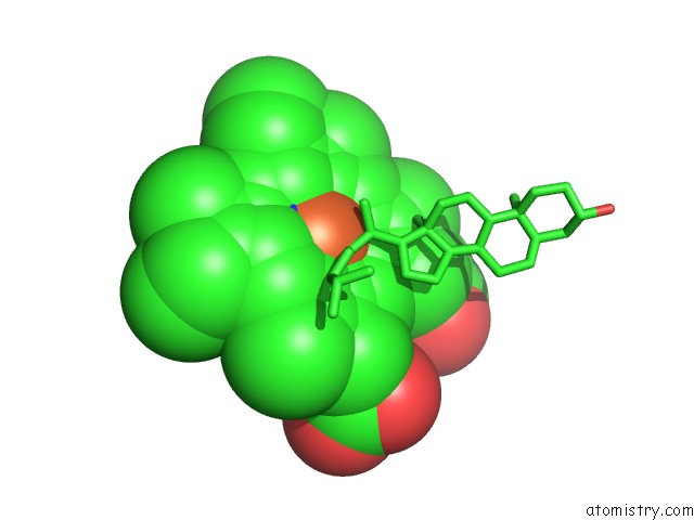
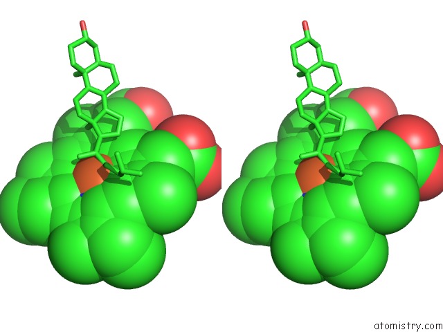
Iron binding site 2 out of 4 in 3mzs
Go back to
Iron binding site 2 out
of 4 in the Crystal Structure of Cytochrome P450 CYP11A1 in Complex with 22- Hydroxy-Cholesterol
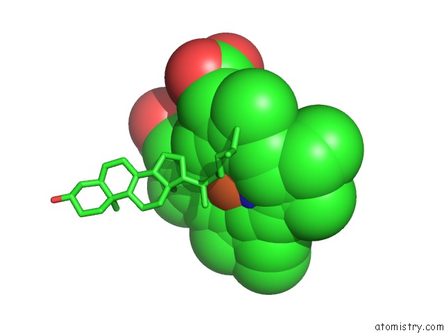
Mono view
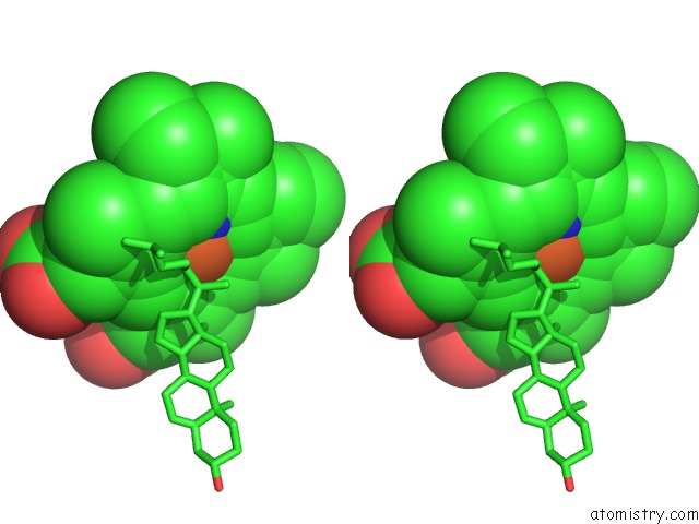
Stereo pair view
Mono view
Stereo pair view
A full contact list of Iron with other atoms in the Fe binding
site number 2 of Crystal Structure of Cytochrome P450 CYP11A1 in Complex with 22- Hydroxy-Cholesterol within 5.0Å range:
|
Iron binding site 3 out of 4 in 3mzs
Go back to
Iron binding site 3 out
of 4 in the Crystal Structure of Cytochrome P450 CYP11A1 in Complex with 22- Hydroxy-Cholesterol
Mono view
Stereo pair view
Mono view
Stereo pair view
A full contact list of Iron with other atoms in the Fe binding
site number 3 of Crystal Structure of Cytochrome P450 CYP11A1 in Complex with 22- Hydroxy-Cholesterol within 5.0Å range:
|
Iron binding site 4 out of 4 in 3mzs
Go back to
Iron binding site 4 out
of 4 in the Crystal Structure of Cytochrome P450 CYP11A1 in Complex with 22- Hydroxy-Cholesterol
Mono view
Stereo pair view
Mono view
Stereo pair view
A full contact list of Iron with other atoms in the Fe binding
site number 4 of Crystal Structure of Cytochrome P450 CYP11A1 in Complex with 22- Hydroxy-Cholesterol within 5.0Å range:
|
Reference:
N.Mast,
A.J.Annalora,
D.T.Lodowski,
K.Palczewski,
C.D.Stout,
I.A.Pikuleva.
Structural Basis For Three-Step Sequential Catalysis By the Cholesterol Side Chain Cleavage Enzyme CYP11A1. J.Biol.Chem. V. 286 5607 2011.
ISSN: ISSN 0021-9258
PubMed: 21159775
DOI: 10.1074/JBC.M110.188433
Page generated: Sun Aug 4 15:47:40 2024
ISSN: ISSN 0021-9258
PubMed: 21159775
DOI: 10.1074/JBC.M110.188433
Last articles
Zn in 9MJ5Zn in 9HNW
Zn in 9G0L
Zn in 9FNE
Zn in 9DZN
Zn in 9E0I
Zn in 9D32
Zn in 9DAK
Zn in 8ZXC
Zn in 8ZUF