Iron »
PDB 3n5s-3na0 »
3n9z »
Iron in PDB 3n9z: Crystal Structure of Human CYP11A1 in Complex with 22- Hydroxycholesterol
Enzymatic activity of Crystal Structure of Human CYP11A1 in Complex with 22- Hydroxycholesterol
All present enzymatic activity of Crystal Structure of Human CYP11A1 in Complex with 22- Hydroxycholesterol:
1.14.15.6;
1.14.15.6;
Protein crystallography data
The structure of Crystal Structure of Human CYP11A1 in Complex with 22- Hydroxycholesterol, PDB code: 3n9z
was solved by
N.V.Strushkevich,
F.Mackenzie,
W.Tempel,
A.Botchkarev,
C.H.Arrowsmith,
A.M.Edwards,
C.Bountra,
J.U.Weigelt,
H.Park,
Structural Genomicsconsortium (Sgc),
with X-Ray Crystallography technique. A brief refinement statistics is given in the table below:
| Resolution Low / High (Å) | 20.00 / 2.17 |
| Space group | P 1 21 1 |
| Cell size a, b, c (Å), α, β, γ (°) | 83.383, 115.095, 86.213, 90.00, 101.82, 90.00 |
| R / Rfree (%) | 21 / 24.3 |
Iron Binding Sites:
The binding sites of Iron atom in the Crystal Structure of Human CYP11A1 in Complex with 22- Hydroxycholesterol
(pdb code 3n9z). This binding sites where shown within
5.0 Angstroms radius around Iron atom.
In total 6 binding sites of Iron where determined in the Crystal Structure of Human CYP11A1 in Complex with 22- Hydroxycholesterol, PDB code: 3n9z:
Jump to Iron binding site number: 1; 2; 3; 4; 5; 6;
In total 6 binding sites of Iron where determined in the Crystal Structure of Human CYP11A1 in Complex with 22- Hydroxycholesterol, PDB code: 3n9z:
Jump to Iron binding site number: 1; 2; 3; 4; 5; 6;
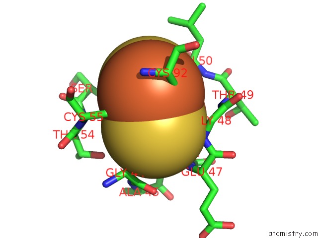
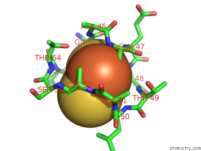
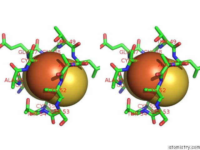
Iron binding site 1 out of 6 in 3n9z
Go back to
Iron binding site 1 out
of 6 in the Crystal Structure of Human CYP11A1 in Complex with 22- Hydroxycholesterol
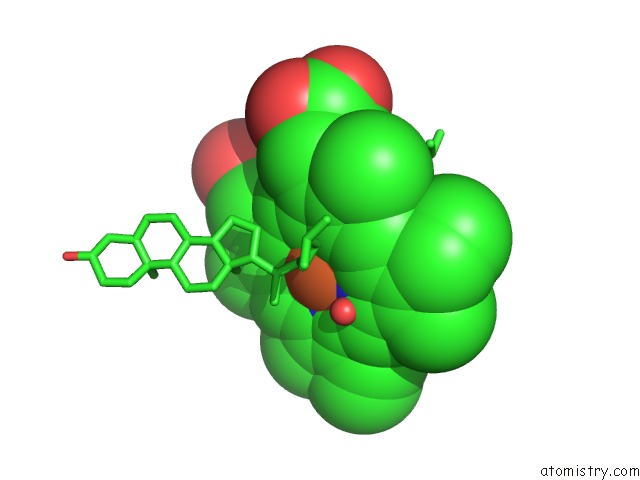
Mono view
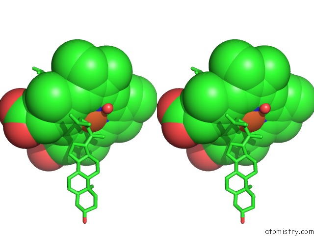
Stereo pair view
Mono view
Stereo pair view
A full contact list of Iron with other atoms in the Fe binding
site number 1 of Crystal Structure of Human CYP11A1 in Complex with 22- Hydroxycholesterol within 5.0Å range:
|
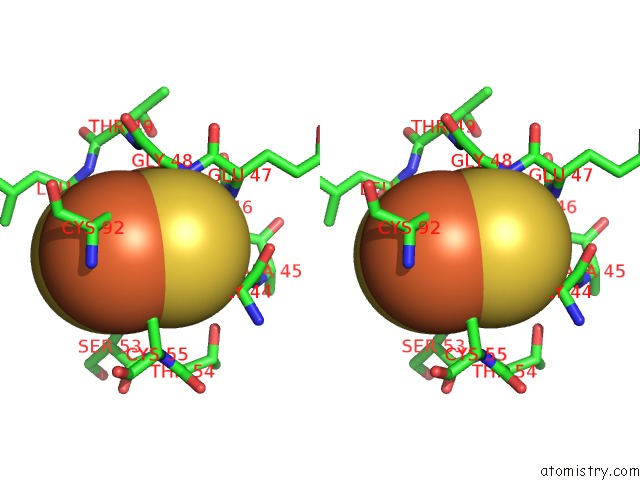
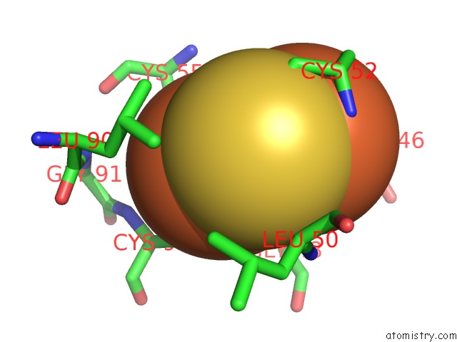
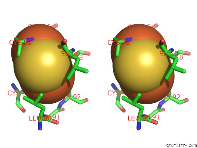
Iron binding site 2 out of 6 in 3n9z
Go back to
Iron binding site 2 out
of 6 in the Crystal Structure of Human CYP11A1 in Complex with 22- Hydroxycholesterol
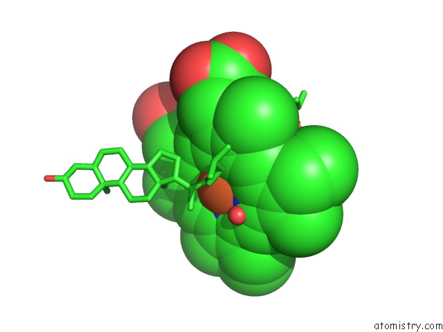
Mono view
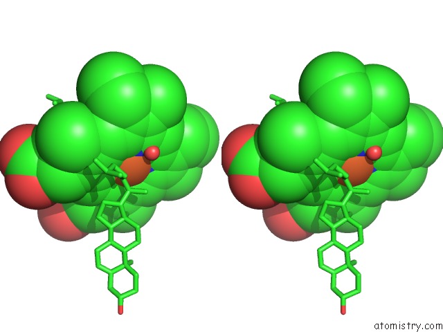
Stereo pair view
Mono view
Stereo pair view
A full contact list of Iron with other atoms in the Fe binding
site number 2 of Crystal Structure of Human CYP11A1 in Complex with 22- Hydroxycholesterol within 5.0Å range:
|
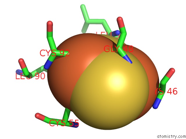
Iron binding site 3 out of 6 in 3n9z
Go back to
Iron binding site 3 out
of 6 in the Crystal Structure of Human CYP11A1 in Complex with 22- Hydroxycholesterol
Mono view
Stereo pair view
Mono view
Stereo pair view
A full contact list of Iron with other atoms in the Fe binding
site number 3 of Crystal Structure of Human CYP11A1 in Complex with 22- Hydroxycholesterol within 5.0Å range:
|
Iron binding site 4 out of 6 in 3n9z
Go back to
Iron binding site 4 out
of 6 in the Crystal Structure of Human CYP11A1 in Complex with 22- Hydroxycholesterol
Mono view
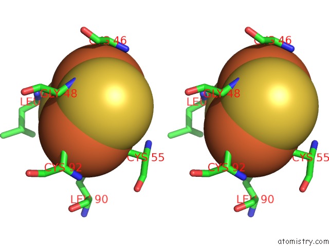
Stereo pair view
Mono view
Stereo pair view
A full contact list of Iron with other atoms in the Fe binding
site number 4 of Crystal Structure of Human CYP11A1 in Complex with 22- Hydroxycholesterol within 5.0Å range:
|
Iron binding site 5 out of 6 in 3n9z
Go back to
Iron binding site 5 out
of 6 in the Crystal Structure of Human CYP11A1 in Complex with 22- Hydroxycholesterol
Mono view
Stereo pair view
Mono view
Stereo pair view
A full contact list of Iron with other atoms in the Fe binding
site number 5 of Crystal Structure of Human CYP11A1 in Complex with 22- Hydroxycholesterol within 5.0Å range:
|
Iron binding site 6 out of 6 in 3n9z
Go back to
Iron binding site 6 out
of 6 in the Crystal Structure of Human CYP11A1 in Complex with 22- Hydroxycholesterol
Mono view
Stereo pair view
Mono view
Stereo pair view
A full contact list of Iron with other atoms in the Fe binding
site number 6 of Crystal Structure of Human CYP11A1 in Complex with 22- Hydroxycholesterol within 5.0Å range:
|
Reference:
N.Strushkevich,
F.Mackenzie,
T.Cherkesova,
I.Grabovec,
S.Usanov,
H.W.Park.
Structural Basis For Pregnenolone Biosynthesis By the Mitochondrial Monooxygenase System. Proc.Natl.Acad.Sci.Usa V. 108 10139 2011.
ISSN: ISSN 0027-8424
PubMed: 21636783
DOI: 10.1073/PNAS.1019441108
Page generated: Sun Aug 4 16:21:55 2024
ISSN: ISSN 0027-8424
PubMed: 21636783
DOI: 10.1073/PNAS.1019441108
Last articles
Fe in 2YXOFe in 2YRS
Fe in 2YXC
Fe in 2YNM
Fe in 2YVJ
Fe in 2YP1
Fe in 2YU2
Fe in 2YU1
Fe in 2YQB
Fe in 2YOO